Hibernom


Hibernomas are rare benign fatty tumors that arise from the vestiges of fetal brown fat and are named “hibernoma” because of their resemblance to the brown fat in hibernating animals .
Epidemiology
Hibernomas usually occur between the ages of 20 and 40 and have a slightly increased incidence in females .
Clinical presentation
They grow slowly and usually present with painless mass effect. Symptoms related to the compression of adjacent structures rarely develop . There are no reports of metastases or malignant transformation .
Pathology
Hibernomas are usually seen in locations where normal brown adipose tissue is present, such as the thigh, shoulder, back, neck, chest, arm (in descending order of frequency), and rarely in the retroperitoneum . Recently, the thigh has been described as a very common location.
Macroscopic appearance
Hibernomas are typically fatty, hypervascular lesions that are grossly similar to a lipoma. They are well-defined, encapsulated, and mobile masses. Their color varies from tan to red-brown, depending on the amount of intracellular lipid. The diameter usually ranges from 5-10 cm, but they may reach up to 20 cm, particularly in the retroperitoneum.
Histology
Upon microscopic examination, the tumors are characterized by cells of various degrees of differentiation. Multivacuolar adipocytes and brown fat cells with granular eosinophilic cytoplasm are interspersed with univacuolar adipocytes. Hypervascularity combined with abundant mitochondria give hibernomas their color .
Morphologic variants of hibernoma in descending order of frequency are
- typical (most common)
- myxoid
- lipoma-like
- spindle cell variants
Genetics
If cytogenic analysis shows an 11q13 translocation, it helps with a diagnosis of hibernoma over liposarcoma .
Radiographic features
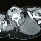
CT
On CT, hibernomas have an attenuation that varies between that of fat and skeletal muscle. They are well circumscribed and may have a peripheral soft tissue attenuation capsule. They enhance slightly and may have a prominent feeding vessel .
MRI
Although they present as brown fat, the imaging characteristics on T1- and T2-weighted images demonstrate high signal intensity but slightly less than that of the subcutaneous fat. On MR imaging, the flow voids of feeding vessels may be identified .
On fat suppression sequences, there may be incomplete fat suppression because of the heterogeneous content of the mass and relatively decreased amount of lipid compared with adipose tissue .
Nuclear medicine
- FDG-PET: FDG avid, although practically this has not been shown to help differentiate from malignancy
Treatment and prognosis
The treatment of hibernomas consists of complete surgical excision. Local recurrence does not occur and there are no reported cases with metastases.
Differential diagnosis
General imaging considerations include:
- liposarcoma
- rhabdomyoma
- resolving hematoma
Siehe auch:
und weiter:


 Assoziationen und Differentialdiagnosen zu Hibernom: