hiläre Lymphadenopathie

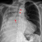
School ager
with a cough. CXR PA and lateral shows massive bilateral hilar lymphadenopathy with the lungs being clear.The diagnosis was tuberculosis.


Sarkoidose in
der Computertomographie: Querschnitt durch den Thorax im Bereich der Aufzweigungen der Bronchien (der Hili) mit vielen vergrößerten Lymphknoten (Pfeile).
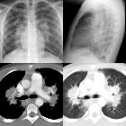
School ager
who 6 years ago had massive hilar lymphadenopathy which eventually resolved with new cough and fever. CXR AP and lateral (above) shows massive bilateral hilar lymphadenopathy and bilateral multiple small lung nodules. Axial CT with contrast of the chest in soft tissue (below left) and lung (below right) windows shows the lymphadenopathy and lung nodules to contain punctate calcifications.The diagnosis was recurrent pulmonary fungal infection in a patient with chronic granulomatous disease.

hiläre Lymphadenopathie
Siehe auch:


 Assoziationen und Differentialdiagnosen zu hiläre Lymphadenopathie: