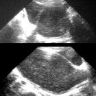
hydrocolpos

Female infant
with pelvic mass. Transverse (above) and sagittal (below) US of the pelvis shows a large cystic structure filled with complex fluid posterior to the bladder which is visible on the right side of the sagittal image. The uterus was identified and was unremarkable and therefore the cystic structure was felt to represent a distended vagina.The diagnosis was hydrocolpos.

Hydrocolpos
• Hydrocolpos - Ganzer Fall bei Radiopaedia

Hydrocolpos
• Congenital hydrocolpos - antenatal ultrasound - Ganzer Fall bei Radiopaedia

Hydrocolpos
• Infantile hydrocolpos - Ganzer Fall bei Radiopaedia
Hydrocolpos is characterized by an expanded fluid filled vaginal cavity. When it is associated with distention of the uterine cavity, the term hydrometrocolpos should then be used. It may present in infancy with a lower abdominal mass, or be delayed till menarche.
Pathology
Etiology
- imperforate hymen (most common)
- vaginal stenosis / transverse vaginal septum
Associations
Radiographic features
Ultrasound
The fluid-filled distended vaginal canal may seen as an anechoic mass between the bladder and the rectum
Differential diagnosis
Consider:
Siehe auch:
- Neoplasien des Ovars
- Rhabdomyosarkom
- pelvic abscess
- Hydrometra
- Hydrometrokolpos
- transverses Vaginalseptum
- haematometrocolpos
- Hämatokolpos
- vaginal obstruction
und weiter:

 Assoziationen und Differentialdiagnosen zu Hydrokolpos:


