Uterus didelphys


Uterus didelphys is a type of Müllerian duct anomaly (class III) where there is a complete duplication of uterine horns as well as duplication of the cervix, with no communication between them.
Epidemiology
Didelphic uteri account for approximately ~8% (range 5-11%) of Müllerian duct anomalies.
Associations
- renal agenesis
- vaginal septum which can include a transverse vaginal septum
- there is a vaginal septum in 75% of cases, and obstruction to one horn is possible from occasional transverse septa
Clinical presentation
Many patients are asymptomatic although some may occasionally experience dyspareunia as a result of the vaginal septum.
Pathology
It results from failed ductal fusion that occurs between the 12 and 16 week of pregnancy and is characterized by two symmetric, widely divergent uterine horns and two cervices. The uterine volume in each duplicated segment is reduced. As with most uterine anatomical anomalies, there is an increased incidence of fertility issues, and Müllerian abnormalities, in general, are over-represented in infertile women. The chance of seeing a pregnancy to term is significantly reduced, down to only 20%, with a third of pregnancies ending in abortion and over half in premature deliveries. Only 40% of pregnancies resulted in living children .
Along with unicornuate uterus, uterus didelphys has the greatest impact on reproductive performance .
Radiographic features
Classically shows two widely spaced uterine corpora, each with a single Fallopian tube. Separate divergent uterine horns with large fundal cleft (as distinct from a septate uterus)
Uterus didelphys should be differentiated from a bicornuate uterus (separation of horns only) and a septate uterus (midline uterine septum).
Hysterosalpingogram (HSG)
HSG demonstrates two separate endocervical canals that open into separate fusiform endometrial cavities, with no communication between the two horns. Each endometrial cavity ends in a solitary fallopian tube.
If the anomaly is associated with an obstructed longitudinal vaginal septum, only one cervical os may be depicted, and it may be cannulated with the endometrial configuration mimicking a unicornuate uterus.
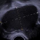
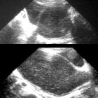
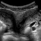
Ultrasound
Separate divergent uterine horns are identified with a large fundal cleft. Endometrial cavities are uniformly separate, with no evidence of communication. Two separate cervices need to be documented.
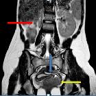
MRI
MR imaging demonstrates two separate uteri with widely divergent apices, two separate cervices, and usually an upper vaginal longitudinal septum. In each uterus, normal uterine zonal anatomy is preserved.
Treatment and prognosis
Complications
infertility
unilateral hydrocolpos/hematocolpos (if a vaginal septum is present)
- endometriosis
See also
Siehe auch:
- Endometriose
- einseitige Nierenagenesie
- Müllerian duct anomaly classification
- Fehlbildungen der Gebärmutter
- Uterus septus
- Uterus bicornis
- Hydrokolpos
- Uterus unicornis
- uterine anomalies
- obstruierte Hemivagina und ipsilaterale renale Anomalie (OHVIRA) Syndrom
- transverses Vaginalseptum
- Hämatokolpos
- Uterus didelphys Schwangerschaft
und weiter:


 Assoziationen und Differentialdiagnosen zu Uterus didelphys: