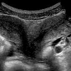
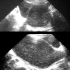
transverse vaginal septum


Transverse vaginal (transvaginal) septum (TVS) is a type of rare congenital uterovaginal anomaly (class II under the Rock and Adam classification).
Epidemiology
It is rare with a frequency of 1 in 70,000 females.
Clinical presentation
In the case of a complete septum, patients commonly present with primary amenorrhea and cyclic pelvic pain.
Rarely diagnosed in neonates or infants, unless the obstruction causes significant hydromucocolpos.
Clinical examination of the vulva is normal if the septum is in the middle/upper vagina. If a membrane is visible, it will not transilluminate, unlike an imperforate hymen .
Pathology
It is a type of vertical fusion defect. A transverse vaginal septum can be either perforate (incomplete) or imperforate (complete) and results from varying degrees of failure in resorption of the tissue between the vaginal plate and the caudal aspect of the fused Müllerian ducts.
The septum is a fibrous membrane of connective tissue with vascular and muscular components. As a result, the functional length of the vagina is reduced.
Associations
While it may occur in isolation, it is often combined with other Müllerian duct anomalies such as uterus didelphys .
Location
It can occur at almost any level of the vagina. Reported prevalence in terms of position include :
- superior vagina (~46%)
- mid-vagina (~40%)
- inferior vagina (~14%)
Radiographic features
Antenatal ultrasound
The diagnosis may be suggested in utero when there is obstruction causing a significant amount of hydrocolpos or mucocolpos.
Treatment and prognosis
Often a surgical excision of the obstructed septum through a perineal approach is performed.
Differential diagnosis
The differential is of congenital absence of the cervix. It is important to identify the cervix on MRI in order to differentiate between a high transverse septum and congenital absence of the cervix. Treatment varies between the two entities, with congenital absence requiring a hysterectomy.
Siehe auch:
und weiter:

 Assoziationen und Differentialdiagnosen zu transverses Vaginalseptum:


