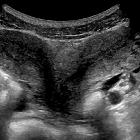
Uterus bicornis


A bicornuate uterus is a type of uterine duplication anomaly. It can be classified as a class IV Mullerian duct anomaly.
Epidemiology
Overall, congenital uterine anomalies occur in ~1.5% of females (range 0.1-3%). Bicornuate uteri are thought to represent ~25% (range 10-39%) of Mullerian duct anomalies .
Clinical presentation
In most cases, a bicornuate uterus is incidentally discovered when the pelvis is imaged. The most common symptomatic presentation is with early pregnancy loss and cervical incompetence . Infertility is not usually a problem with this type of malformation because implantation of the embryo is not impaired.
Associations
- a longitudinal vaginal septum may be present in ~25% of cases
- as with other Mullerian duct anomalies, abnormalities of the renal tract may also be present
Pathology
A bicornuate uterus results from abnormal development of the paramesonephric ducts. There is a partial failure of fusion of the ducts, resulting in a uterus divided into two horns.
Subtypes
A bicornuate uterus is divided according to the involvement of the cervical canal:
- bicornuate bicollis: two cervical canals; central myometrium extends to the external cervical os
- bicornuate unicollis: one cervical canal; central myometrium extends to the internal cervical os
Radiographic features
General
The preferred methods of imaging uterine anomalies are ultrasound, hysterosalpingogram or MRI. The external uterine contour is concave or heart-shaped, and the uterine horns are widely divergent. The fundal cleft is typically more than 1 cm deep and the intercornual distance is widened.
The uterus is seen as comprising caudally fused symmetric uterine cavities with some degree of communication between the two cavities (usually at the uterine isthmus). Although not a specific finding, the angle between the horns of the bicornuate uterus is usually more than 105° .
Hysterosalpingogram (HSG)
A divided uterus can be seen, but it is difficult to differentiate between septate and bicornuate anomalies since the uterine fundal contour is not visible . Accuracy of hysterosalpingogram alone is only 55% in differentiation between both entities. An angle of less than 75° between the uterine horns is suggestive of a septate uterus while an angle of more than 105° is more consistent with a bicornuate uterus.
MRI
May help confirm anatomy by showing a deep (>1 cm) fundal cleft in the outer uterine contour and an intercornual distance of >4 cm. The uterus demonstrates normal uterine zonal anatomy.
Treatment and prognosis
Surgical intervention is usually not indicated in the absence of reproductive difficulties.
In women with a history of recurrent pregnancy loss and in whom no other infertility issues have been identified a Strassman metroplasty can be considered.
In patients with cervical incompetence, placement of a cervical cerclage may increase fetal survival rates . Indeed the association between cervical incompetence and a bicornuate uterus is so high, that prophylactic cerclage may be appropriate in some instances.
Differential diagnosis
- uterus didelphys: complete failure of fusion occurs during the development of the paramesonephric ducts with duplication of the uterus, cervix and vagina
- septate uterus: has a normal fundal contour but is characterized by a persistent longitudinal septum that partially divides the uterine cavity
Practical points
Septate uterus increases the risk of early pregnancy loss and hysteroscopic intervention to resect the septum is sometimes pursued. In this situation, differentiation between a septate uterus or a bicornuate uterus is critical. This is mostly a problem with HSG preoperative evaluation. Attempted resection of a bicornuate uterus "septum" leads to a poor outcome.
Siehe auch:
- Uterus didelphys
- Fehlbildungen der Gebärmutter
- cervical incompetence
- Uterus septus
- Müller-Gang
- Duplikatur des Uterus
- Schwangerschaft bei Uterus bicornis
- class IV Mullerian duct anomaly
und weiter:

 Assoziationen und Differentialdiagnosen zu Uterus bicornis:


