Hydropneumothorax

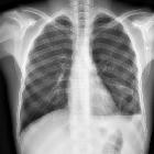
Seropneumothorax
rechts: Unterlappen und Mittellappen scheinen im Pleuraerguss. Die Lungenspitze liegt dem thorakalen Apex nicht an.

Seropneumothorax
nach Rippenfrakturen sekundär aufgetreten. Basaler Spiegel.

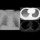
Teenager in a
motor vehicle accident with chest painCXR AP shows a widened mediastinum and a small right-sided pneumothorax. Axial CT with contrast of the chest with lung (above) and bone (below) windows shows air in the pleural space anterior to the right lung and fluid in the pleural space posterior to the right lung as well as airspace disease in the right lower lobe and a cortical disruption through one of the right ribs posteriorly.The diagnosis was a right hydropneumothorax, right pulmonary contusion, and right posterior rib fracture. Angiogram was negative for aortic injury.

Hydropneumothorax
• Hydropneumothorax - Ganzer Fall bei Radiopaedia

Hydropneumothorax
• Traumatic hydropneumothorax - Ganzer Fall bei Radiopaedia

Hydropneumothorax
• Hydropneumothorax - Ganzer Fall bei Radiopaedia

Hydropneumothorax
• Hydropneumothorax - Ganzer Fall bei Radiopaedia

Hydropneumothorax
• Hydropneumothorax - Ganzer Fall bei Radiopaedia

Hydropneumothorax
• Hydropneumothorax, pneumomediastinum, pneumoperitoneum and pneumoretroperitoneum - Ganzer Fall bei Radiopaedia

Hydropneumothorax
• Osteosarcoma metastases - Ganzer Fall bei Radiopaedia

Hydropneumothorax
• Hydropneumothorax - Ganzer Fall bei Radiopaedia

Hydropneumothorax
• Hydropneumothorax - Ganzer Fall bei Radiopaedia
A hydropneumothorax (plural: hydropneumothoraces) (or less commonly pneumohydrothorax (plural: pneumohydrothoraces)) is the term given to the concurrent presence of a pneumothorax and pleural effusion (i.e. hydrothorax) (i.e. gas and fluid) in the pleural space.
Pathology
It may arise in various situations which include
- thoracentesis
- thoracic trauma
- bronchopleural fistula
- esophagopleural fistula
Radiographic features
Plain radiograph
On an erect chest radiograph, recognition of hydropneumothorax can be rather easy - and is classically shown as a gas-fluid level. On the supine radiograph, this may be more challenging where a sharp pleural line is bordered by increased opacity lateral to it within the pleural space may sometimes suggest towards the diagnosis .
Ultrasound
Sonographic findings include ;
- hydro-point sign
- describes the interface of a pleural line which lacks lung sliding and an anechoic collection bounded inferiorly by the diaphragm
- coexistence of the sonographic signs characteristic of a pneumothorax and a pleural effusion
- a lung point sign may also be found
- describes the interface of a pleural line which lacks lung sliding and an anechoic collection bounded inferiorly by the diaphragm
Differential diagnosis
For a gas-fluid level on plain radiograph consider:
- pyopneumothorax
- hemopneumothorax
- pulmonary abscess
- traumatic pneumatocele
- soft tissue abscess
Siehe auch:
und weiter:

 Assoziationen und Differentialdiagnosen zu Seropneumothorax:


