hypoxic-ischemic encephalopathy
Newborn who
experienced prolonged labor. Axial (above), coronal (below left) and sagittal (below right) CT without contrast of the brain show a large low density fluid collection in the subcutaneous tissues of the scalp that crosses sutures and is seen to surround the skull on the coronal view. Intracranially, there is diffuse loss of gray matter-white matter differentiation secondary to diffuse cerebral edema.The diagnosis was caput succedaneum in a patient with hypoxic ischemic encephalopathy.

Hypoxic-ischemic
encephalopathy (adults and children) • Hypoxic-ischemic brain injury - Ganzer Fall bei Radiopaedia

Hypoxic-ischemic
encephalopathy (adults and children) • Hypoxic-ischemic brain damage - Ganzer Fall bei Radiopaedia

Hypoxic-ischemic
encephalopathy (adults and children) • Global hypoxic ischemic brain injury - Ganzer Fall bei Radiopaedia

Hypoxic-ischemic
encephalopathy (adults and children) • Near drowning - Ganzer Fall bei Radiopaedia

Hypoxic-ischemic
encephalopathy (adults and children) • Global hypoxic ischemic encephalopathy - Ganzer Fall bei Radiopaedia

Hypoxic-ischemic
encephalopathy (adults and children) • Hypoxic encephalopathy - Ganzer Fall bei Radiopaedia

Hypoxic-ischemic
encephalopathy (adults and children) • Global hypoxic injury post cardiac arrest - Ganzer Fall bei Radiopaedia

Pseudosubarachnoid
hemorrhage • Global hypoxic-ischemic brain injury - Ganzer Fall bei Radiopaedia

Hypoxic-ischemic
encephalopathy (adults and children) • Hypoxic ischemic encephalpathy - Ganzer Fall bei Radiopaedia

Hypoxic-ischemic
encephalopathy (adults and children) • Global hypoxic brain injury - Ganzer Fall bei Radiopaedia

Hypoxic-ischemic
encephalopathy (adults and children) • Brain cortical laminar necrosis - Ganzer Fall bei Radiopaedia

Hypoxic-ischemic
encephalopathy (adults and children) • Hypoxic-ischemic brain injury - Ganzer Fall bei Radiopaedia

Hypoxic-ischemic
encephalopathy (adults and children) • Global cerebral ischemia - Ganzer Fall bei Radiopaedia

Hypoxic-ischemic
encephalopathy (adults and children) • Global hypoxic injury - Ganzer Fall bei Radiopaedia

Susceptibility
weighted imaging • Hypoxic-ischemic encephalopathy and hyperoxygenation - Ganzer Fall bei Radiopaedia
Hypoxic-ischemic
encephalopathy (adults and children) • Basal ganglia chronic hypoxic ischemic disease - Ganzer Fall bei Radiopaedia

Basal ganglia
T2 hyperintensity • Basal ganglia chronic hypoxic ischemic disease - Ganzer Fall bei Radiopaedia

Hypoxic-ischemic
encephalopathy (adults and children) • CNS cryptococcosis - Ganzer Fall bei Radiopaedia

Hypoxic-ischemic
encephalopathy (adults and children) • Hypoxic ischemic encephalopathy - Ganzer Fall bei Radiopaedia

Pseudosubarachnoid
hemorrhage • Hypoxic-ischemic brain injury - Ganzer Fall bei Radiopaedia

Anoxic brain
injury • Global hypoxic ischemic encephalopathy - Ganzer Fall bei Radiopaedia

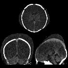
Newborn with
failure to progress during delivery due to dystocia who is now apneic. Axial CT without contrast of the brain shows a cresenteric high-density fluid collection in the subcutaneous tissues of the right scalp that crosses suture lines and a cresenteric low-density fluid collection in the subcutaneous tissues of the left scalp that crosses suture lines. Intracranially, there is diffuse loss of gray matter-white matter differentiation secondary to diffuse cerebral edema.The diagnosis was a left caput succedaneum and a right subgaleal hematoma in a patient with hypoxic ischemic encephalopathy.

Hypoxic
ischaemic encephalopathy. T2 hyperintensity seen in both cerebellar hemispheres.

Anoxic brain
injury following a hanging. Note the loss of grey white matter differentiation and small ventricles due to brain swelling.

This image
is part of a series which can be scrolled interactively with the mousewheel or mouse dragging. This is done by using Template:Imagestack. The series is found in the category Profound hypoxia case 001. Computertomographie des Schädels bei einem Patienten nach generalisierter Hypoxie: Besonders die stoffwechselintensiven und somit hypoxieempfindlichen Regionen wie Globus pallidum, Hippocampus und auch die Hirnschenkel sind betroffen. Auch die Mark-Rinden-Differenzierung ist deutlich vermindert.

Computertomographie
des Schädels bei einem jungen Patienten nach generalisierter Hypoxie: Die stoffwechselintensiven und somit hypoxieempfindlichen Regionen hier vor allem Globus pallidum sind betroffen und somit deutlich hypodens.

Schwerer
hypoxischer Hirnschaden mit aufgehobener Mark-Rinden-Differenzierung. Computertomographie axial.


Hypoxic
ischaemic encephalopathy. Diffusion restriction seen in both cerebellar hemispheres.

Hypoxic
ischaemic encephalopathy. Diffusion restriction seen in both cerebral hemispheres, basal ganglia and thalami.

Hypoxic
ischaemic encephalopathy. Both cerebral hemispheres, basal ganglia and thalami show low ADC map values.

Hypoxic-ischemic
encephalopathy (adults and children) • Anoxic brain injury - Ganzer Fall bei Radiopaedia

White
cerebellum sign • Diffuse hypoxic ischemic brain injury - Ganzer Fall bei Radiopaedia

Hypoxic
ischaemic encephalopathy. FLAIR Hyperintensity seen in both cerebellar hemispheres.

Acute carbon
monoxide poisoning and hypoxic-ischaemic brain injury. Diffuse hypoattenuation of gray matter in cerebral cortex and effacement of cerebral sulci, obliteration of the fourth ventricle and the basal cisterns.

Acute carbon
monoxide poisoning and hypoxic-ischaemic brain injury. Bilateral basal ganglia low density (lenticular nucleus, head of the caudate nucleus and thalamus) and diffuse hypo-attenuation of gray matter in cerebral cortex with relative preservation of white matter.
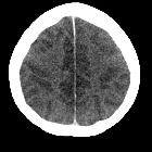
Acute carbon
monoxide poisoning and hypoxic-ischaemic brain injury. Diffuse hypoattenuation of gray matter in cerebral cortex with relative preservation of white matter and effacement of cerebral sulci.

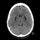
Infant who is
status post cardiac arrest. Axial CT without contrast of the brain shows decreased density of the cerebrum when compared to the density of the cerebellum. There is also loss of gray matter-white matter differentiation and effacement of the cerebral sulci and the basal cisterns. There is linear increased density present along the entire falx.The diagnosis was hypoxic ischemic encephalopathy and interhemispheric subdural hematoma in a child abuse patient.

Newborn with
a nuchal cord. Axial CT without contrast of the brain (above) shows diffuse cerebral edema causing loss of the gray matter white matter junction. Axial T1 without contrast (middle left), FLAIR (middle middle) and T2 (middle right) MRI several days later show developing laminar necrosis. Diffusion weighted imaging (below) shows diffusion restriction throughout the gray matter, white matter, and basal ganglia.The diagnosis was severe hypoxic ischemic encephalopathy.

Anoxic brain
injury • Anoxic brain injury - Ganzer Fall bei Radiopaedia

Anoxic brain
injury • Anoxic brain injury - Ganzer Fall bei Radiopaedia

White
cerebellum sign • Anoxic brain injury - Ganzer Fall bei Radiopaedia

Hypoxic-ischemic
encephalopathy (adults and children) • Hypoxic ischemic encephalopathy - Ganzer Fall bei Radiopaedia
hypoxischer Hirnschaden
hypoxic-ischemic encephalopathy
Siehe auch:
- kortikale laminäre Nekrose
- Pseudosubarachnoidalblutung
- reversal sign
- Kohlenmonoxidintoxikation
- neonatal hypoxic-ischaemic encephalopathy
- hypoxic-ischaemic injury in older children and adults
- global cerebral ischemia
- Globus pallidum hypodens
und weiter:
- hypoxischer Hirnschaden
- Ischämischer Schlaganfall
- intrakranielle Thrombektomie
- dense cerebellum sign
- Enzephalopathie
- fetal hypoxia
- pseudosubarachnoid sign
- MR spectroscopy of hypoxic-ischemic injury
- hypoxic brain damage T2
- Hirntoddiagnostik
- CT in hanging
- hypoxic brain injury with pseudosubarachnoid haemorrhage
- hypoxic brain damage MRI
- hypodense Basalganglien


 Assoziationen und Differentialdiagnosen zu hypoxischer Hirnschaden:
