incisive canal cyst


Incisive canal cysts, also known as nasopalatine duct cysts (NPDC), are developmental, non-neoplastic cysts arising from degeneration of nasopalatine ducts. These ducts usually regress in fetal life. The persistence of ductal epithelium leads to formation of cyst.
It is considered the most common non-odontogenic cyst and develops only in the midline anterior maxilla.
Epidemiology
They most commonly occur in 4 to 6 decades, have a male predominance and affect ~1% of the population.
Clinical presentation
Most patients are symptomatic. They present as swelling of anterior hard palate, sometimes associated with pain and drainage. They must usually be larger than 0.6 cm to distinguish cyst from incisive foramen, which is a normal anatomical feature.
Pathology
Cyst formation is due to spontaneous cystic degeneration of residual ductal epithelium. The vast majority of cases contain non-keratinized stratified squamous epithelium alone or in combination with other epithelia histologically. Approximately 30% cases contain respiratory epithelium .
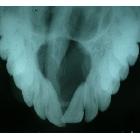
Radiographic features
They are seen as a solitary well-defined, oval or round unilocular radiolucency, between central incisors, >0.6 cm in diameter. They may appear “heart-shaped” if the anterior nasal spine superimposed. Root resorption and tooth displacement may be present.
MRI
Usually shows homogeneous high signal intensity on both on T1 and T2, although most cysts of maxillofacial regions demonstrate low to intermediate signal intensity on T1 weighted images .
Treatment and prognosis
Enucleation is usually curative, and recurrence is rare. Histological confirmation is recommended.
Differential diagnosis
General imaging differential considerations include:
- periapical granuloma/periapical cyst
- schwannoma in the incisive canal region
- nasolabial cyst
See also
Siehe auch:


 Assoziationen und Differentialdiagnosen zu Zyste Canalis incisivus:


