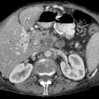
inflammatorischer Pankreaspseudotumor


Mass-forming chronic pancreatitis occurs in around 30% of cases of chronic pancreatitis, where a mass or a focal enlargement of the pancreas is usually seen on imaging. In many instances, it poses a challenge as the epidemiology and imaging appearances overlap those of pancreatic adenocarcinoma.
Epidemiology
- ~ 30% of patients with chronic pancreatitis
- ~ 70% of mass-forming chronic pancreatitis manifest in the pancreatic head
Clinical presentation
Symptoms and risk factors tend to overlap with those of malignancy, including abdominal pain, weight loss, nausea, and jaundice .
Radiographic features
Generally, it manifests with a focal enlargement or a mass-like lesion of the pancreas, which is hypoenhancing . It may be associated with main pancreatic duct and common bile duct dilatation when localized at the pancreatic head, but they commonly taper smoothly and are not completely obstructed (cf. abrupt cutoff in pancreatic adenocarcinoma) - duct penetrating sign.
CT
- hypoattenuating and hypoenhancing
- features that help supporting an inflammatory process over malignancy :
MRI
- T1: hypo to isointense
- T2: iso to hyperintense
- C+ (Gd): variable pattern of enhancement on dynamic phases, usually with loss of the early/arterial phase pancreatic enhancement
Treatment and prognosis
Endoscopic ultrasound (EUS) and fine-needle aspiration (FNA) need to be considered in the equivocal imaging cases.
Studies have shown that ~5%-10% of resected pancreatic head carcinomas are confirmed to represent an inflammatory mass .
Differential diagnosis
- pancreatic adenocarcinoma
- peripheral displacement of chronic pancreatitis calcifications may suggest underlying malignancy
- double duct sign
- soft tissue encasement of adjacent vasculature
- autoimmune pancreatitis
- paraduodenal pancreatitis
- non-functioning pancreatic neuroendocrine tumor
- solid pseudopapillary tumor
- pancreatic metastasis
Siehe auch:

 Assoziationen und Differentialdiagnosen zu inflammatorischer Pankreaspseudotumor:

