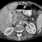
paraduodenal pancreatitis


Paraduodenal pancreatitis is an uncommon type of focal chronic pancreatitis affecting the groove between the head of the pancreas, the duodenum and the common bile duct.
Terminology
The following entities with which it shares clinicopathological features are unified by this term and should no longer be used :
- cystic dystrophy of heterotopic pancreas
- pancreatic hamartoma of duodenum
- myoadenomatosis
- duodenal wall cyst
- groove pancreatitis
Epidemiology
The epidemiology of paraduodenal pancreatitis is similar to that of run-of-the-mill chronic pancreatitis.
This rare entity is predominantly seen in males with a history of alcohol abuse. Peak incidence is roughly 40-50 years old .
Clinical presentation
The clinical presentation of paraduodenal pancreatitis is dominated by symptoms resulting from marked duodenal stenosis and impaired motility with patients presenting with nausea and vomiting.
Symptoms comprise:
- recurrent episodes of upper abdominal pain and nausea, typically aggravated by fat or protein-rich meals
- weight loss from poor nourishment
Possible signs include:
- slight elevation of serum markers
- pancreatic enzymes (amylase and lipase)
- cholestatic liver enzymes gamma-glutamyltransferase (GGT) and alkaline phosphatase (ALP)
- usually unremarkable level of tumor markers, e.g.
Jaundice is a rare finding, despite the frequent involvement of the common bile duct in terms of tubular stenosis.
Pathology
The pathogenesis of paraduodenal pancreatitis remains controversial.
The hallmark of disease is the presence of scar tissue with fibrosis in the pancreaticoduodenal groove or in the groove and the superior portion of the pancreatic head (in the pure and segmental forms of the disease, respectively).
The duodenum is always involved by a chronic inflammatory process, with scar tissue in the wall leading to fibrosis and various levels of stenosis.
Etiology
Paraduodenal pancreatitis has been suggested to occur via increased viscosity of the pancreatic juice caused by alcohol, thus predisposing to crystal formation and increased production of protein, resulting in stone formation. Impeding the minor papilla either pathoanatomically or functionally this process incapacitates exocrine pancreatic function.
The presence of affected pancreatic elements in the duodenal wall leads to inflammatory injury of the descending (second) part of the duodenum in the pancreatic groove. Changes may be cystic and/or stenotic .
Microscopic appearance
Histopathological features have been demonstrated to include :
- dilated pancreatic ducts
- duodenal wall pseudocysts
- granulation tissue containing fused macrophages of foreign-body type
- hyperplasia of Brunner glands
- heterotopic pancreatic acinar tissue interspersed by dense myofibroblastic and neural proliferation
- involvement of the pancreaticoduodenal groove by fibrotic inflammatory changes
Radiographic features
Paraduodenal pancreatitis is usually classified into two forms, pure and segmental:
- pure form: affects exclusively the groove
- segmental form: extends to the pancreatic head despite a clear predominance in the groove
Ultrasound
Transabdominal ultrasound
May demonstrate :
- thickening of the second part of the duodenum
- possibly in continuity to a hypoechoic mass near the pancreatic head
- additional findings reflecting the background pathologic processes
- chronic calcific pancreatitis
- mild to moderate dilatation of pancreatic duct and common bile duct dilatation, possibly mimicking the "double duct sign"
Endoscopic ultrasound (EUS)
May be preferred over transabdominal imaging as it more readily demonstrates the expected pathological changes comprising of:
- circumscript hypoechoic change between the duodenal wall and the pancreatic parenchyma
- thickening of duodenal wall and associated stenosis
- pancreatic calcifications and pseudocysts
- stenotic changes of the common bile duct
- dilatation of the pancreatic duct
EUS may also allow for guided needle tissue samples .
CT
May depict pathological changes accurately. The following findings have been described in the literature:
- cystic thickening of the duodenal wall with or without duodenal stenosis
- fibrous tissue within the pancreaticoduodenal groove (may show late enhancement after contrast administration)
- common bile duct dilatation
MRI
The most characteristic finding on MRI is a sheetlike mass between the head of the pancreas and the duodenum .
Signal characteristics include:
- T1: hypointense to pancreatic parenchyma
- T2: hypo-, iso-, or slightly hyper-intense
Associated findings include:
- inflammatory changes of the pancreas
- duodenal stenosis
- local cysts
- regular tapering of the pancreatic and common bile ducts
- widening of the space between the distal pancreatic and common bile ducts, and duodenal lumen at MRCP
- banana-shaped gallbladder
Treatment and prognosis
Conservative treatment options include analgesics, pancreatic rest and abstinence from alcohol. Symptom relief by these measures is frequently not permanent, as they are unable to stop the underlying background pathologic changes.
Organ-sparing surgery with modified Whipple procedure as either pylorus-preserving pancreaticoduodenectomy or Frey procedure have been demonstrated to result in good and lasting pain relief together with improvement in quality of life .
History and etymology
It was first described by V Becker (German physician) in 1973 .
The term “groove pancreatitis” appeared later, coined in 1982 by Stolte et al, members of Becker's group. It is the direct translation of the original term "Rinnenpankreatitis".
A distinction between "pure groove pancreatitis" and "segmental groove pancreatitis" was also reported by the Becker group :
- “pure groove pancreatitis”: scarring limited to the groove area
and, - “segmental groove pancreatitis,” involvement of the pancreatic head
Differential diagnosis
- pancreatic adenocarcinoma of the head of the pancreas is the most relevant differential diagnosis of paraduodenal pancreatitis (particularly in its segmental form). Needle tissue (FNA) samples may be a challenge for the interpreting pathologist
- duodenal adenocarcinoma mainly when they appear as focal thickening of the medial duodenal wall could be hard to differentiate from paraduodenal pancreatitis
- ampullary carcinoma
- acute pancreatitis involving the pancreaticoduodenal groove

 Assoziationen und Differentialdiagnosen zu paraduodenale Pankreatitis:


