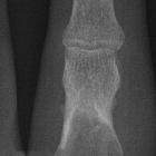
intraosseous ganglia

MRI
characteristics of cysts and “cyst-like” lesions in and around the knee: what the radiologist needs to know. Intraosseous ganglion cyst. The axial (a) and coronal (b) fat saturated proton density weighted images demonstrate a solitary, unilocular cystic lesion (arrows). Note the sclerotic rim (arrowheads) and mild reactive bone oedema (asterisk)

Intraosseous
ganglion • Intraosseous ganglion cyst - Ganzer Fall bei Radiopaedia

Intraosseous
ganglion • Intraosseous ganglion - Ganzer Fall bei Radiopaedia

Intraosseous
ganglion • Intraosseous ganglion of the knee - Ganzer Fall bei Radiopaedia

Intraosseous
ganglion • Intraosseous ganglion cyst - Ganzer Fall bei Radiopaedia

Spectrum of
MRI features of ganglion and synovial cysts. Intraosseous ganglion cyst of the tibia incidentally depicted in a 40-year-old man who underwent an MRI scan due to intermittent, subacute non-specific knee pain. Sagittal FS PD-WI shows a metaepiphyseal, large, multiloculated cystic lesion of the tibia, which communicates with the articular surface through a thin stalk (arrow) extending into the interspinous region, close to the anterior cruciate ligament tibial insertion. Tb, tibia; ACL, anterior cruciate ligament
An intraosseous ganglion (plural: ganglia) is a benign subchondral radiolucent lesion without degenerative arthritis.
Epidemiology
Tends to occur in middle age.
Clinical presentation
Patients may have mild localized pain.
Pathology
They are uni-/multilocular cysts surrounded by a fibrous lining, containing gelatinous material.
Origin
Location
Common locations are:
- epiphyses of long bones (medial malleolus, femoral head, proximal tibia, carpal bones)
- subarticular flat bone (acetabulum)
Radiographic features
Plain radiograph
Typically well-demarcated solitary lytic lesion, with a sclerotic margin. No communication with joint can be demonstrated.
MRI
- solitary, unilocular or multilocular
- usually sclerotic rim is present
Bone scan
Bone scans demonstrate increased radiotracer uptake (in 10%).
Differential diagnosis
- post-traumatic/degenerative cyst
See also
Siehe auch:
- nicht ossifizierendes Fibrom
- Enchondrom
- intraossäre Zyste mit Lufteinschluss
- subchondrale Zysten
- Ganglion (Überbein)
- Riesenzelltumor des Knochens
- Knochenläsionen der Epiphyse
- Geode vs intraossäres Ganglion
und weiter:

 Assoziationen und Differentialdiagnosen zu intraossäres Ganglion:


