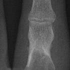
Riesenzelltumor des Beckenknochens

Transarterial
embolization (TAE) of sacral giant cell Tumor (GCT) using spherical parmanent embolic material superabsorbant polymer microsphere (SAP-MS). A 68-year-old female with GCT in the sacrum (Case 2). A. On CT, a large osteolytic mass occupied the sacrum (white arrows). B. On CT of same level as Figure 1A about 10 years after initial TAE, the size of the mass decreased and obvious reossification is shown (white arrows).

Transarterial
embolization (TAE) of sacral giant cell Tumor (GCT) using spherical parmanent embolic material superabsorbant polymer microsphere (SAP-MS). A 40-year-old female with GCT in the sacrum (Case 3). A. On contrast enhanced fat supressed T1-weighted image of MRI, the homogenous enhanced mass occupied the sacrum (white arrows). B. On digital sabtraction pelvic angiogram, the hypervascular mass is shown in the sacrum (arrow). C. On digital sabtraction pelvic angiogram after TAE of bilateral lateral sacral arteries and middle sacral artery, the tumor stain is diminished (arrow). D. On contrast enhanced fat supressed T1-weighted image of MRI about 14 months after initial TAE, the enhanced pattern becomes inhomogenous compared with Figure 2A, which shows what appears to be a necrotic effect (white arrows).

Pelvic bone
giant cell tumour. CT images help confirming lack of internal matrix mineralization.

Pelvic bone
giant cell tumour. T2-weighted image: The lesion shows low to intermediate signal intensity.

Pelvic bone
giant cell tumour. T1-weighted image: The lesion shows low to intermediate signal intensity.

Pelvic bone
giant cell tumour. Before Gadolinium-Based Contrast Media administration

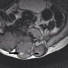
Pelvic bone
giant cell tumour. After Gadolinium-Based Contrast Media administration: Tumour shows solid enhancement.
Giant cell
tumour of sacrum. Axial T2WI, high signal area within soft tissue representing necrotic /cystic component (arrow).
Riesenzelltumor des Beckenknochens
Siehe auch:
- nicht ossifizierendes Fibrom
- Enchondrom
- Aneurysmatische Knochenzyste
- Chondrosarkom
- intraossäres Ganglion
- Multiples Myelom
- primärer Hyperparathyreoidismus
- Chondroblastom
- Riesenzelltumor
- Chondromyxoidfibrom
- desmoplastisches Fibrom
- Riesenzelltumor der Sehnenscheiden
- Riesenzelltumor des Knochens
- Ostitis fibrosa cystica
- Riesenzelltumor des Sakrums
- Osteoklastom
- Beckentumoren
- Metastasen des Beckens


 Assoziationen und Differentialdiagnosen zu Riesenzelltumor des Beckenknochens: