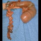
jejunal atresia


Jejunal atresia is a congenital anomaly characterized by obliteration of the lumen of the jejunum. The site of the atresia can be anywhere from the ligament of Treitz to the jejunoileal junction. There can be more than one atretic segment.
This article will focus on jejunal atresia alone but bear in mind that some cases correspond to jejunoileal atresia and show a mixed pattern, including the ones discussed in the ileal atresia article.
Epidemiology
Jejunal atresia has an incidence of about 1:1,000 live births and is more common than duodenal atresia.
Clinical presentation
Neonates typically present with abdominal distension and bilious vomiting within the first 24 hours of birth. These symptoms, however, do not allow for differentiation from a duodenal atresia.
Pathology
The etiology is thought to be from an in-utero ischemic event.
Radiographic features
Plain radiograph
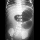
The classic radiographic sign of jejunal atresia is that of a triple bubble appearance for a proximal obstruction; it is equivalent to the double bubble sign of duodenal appearance plus a third bubble is caused by filling and distention of the jejunum by air.
There can be multiple dilated small bowel loops proximal to the atresia and the number of dilated loops increase as point of atresia becomes more distal.
Fluoroscopy
Contrast enema typically shows micro colon (small unused colon)
Antenatal ultrasound
- may show dilated proximal bowel loops, often greater than 7 mm
- may show evidence of an in utero bowel perforation
- there may be polyhydramnios, especially in cases where the atresia is proximal
Differential diagnosis
On plain radiograph consider:
- malrotation with midgut volvulus
On contrast enema consider:
See also
Siehe auch:
und weiter:

 Assoziationen und Differentialdiagnosen zu Jejunalatresie:


