Kontusionsblutung


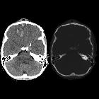
Cerebral hemorrhagic contusions are a type of intracerebral hemorrhage and are common in the setting of significant head injury. They are usually characterized on CT as hyperdense foci in the frontal lobes adjacent to the floor of the anterior cranial fossa and in the temporal poles.
Epidemiology
Contusions, by definition, result from head trauma and are thus seen more frequently in young males. Typical causes include motor vehicle accidents or situations in which the head strikes the ground.
Pathology
Most contusions represent the brain coming to a sudden stop against the inner surface of the skull (contrecoup) accentuated by the natural contours of the skull (see below).
Associations
- intraventricular hemorrhage in ~2.5%
Radiographic features
Cerebral contusions can occur anywhere, but have a predilection for certain locations, as a result of the direction of the head strike and the intrinsic shape of the skull cavity.
- anterior cranial fossa floor
- temporal pole
- coup and contrecoup pattern
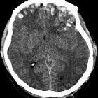
Typically cortical contusions become more apparent on follow-up imaging due to further bleed or surrounding edema. Hence on follow-up CT scans in the first couple of days after trauma, one may detect the increase in number and size of the lesions but the patient may not show any clinical deterioration.
Furthermore, the appearance of contusions will vary according to when they are imaged. Typically they mature over some weeks, initially appearing as merely hemorrhagic foci, followed by the development of surrounding edema, before gradually fading away leaving behind more or less distinct areas of gliosis.
CT
In most hospitals, CT is usually the first and often the only investigation used to assess cerebral contusions. Sensitivity to detect intracerebral hemorrhage on CT scans is virtually 100%.
Hounsfield units (HU) of blood are dependent on protein concentration (i.e. hemoglobin) and hematocrit.
With a hematocrit of 45%, the density of whole blood is 45-65 HU while the grey matter is 37-41 HU and white matter is 30-34 HU. So blood should be hyperdense in comparison to grey or white matter.
Of note, in anemic patients (i.e. hemoglobin <8-10 g/dL) blood may appear isodense in acute bleeding.
Contusions vary in size and can appear as small petechial foci of hyperdensity/hemorrhages involving the grey matter and subcortical white matter or large cortical/subcortical bleed.
MRI
Although not often used merely for the assessment of superficial contusions, MRI is far more sensitive to small contusions, especially on T2* sequences, e.g. SWI.
- T1: high signal intensity
- T2*: low signal intensity
- SWI: low signal intensity
Signal behavior is strongly dependent on sequence and time since the bleeding started.
Differential diagnosis
- diffuse axonal injury
- cavernoma
- cerebral contusions undergo expected evolution of blood products whereas cavernoma stay stable or re-bleed
- look for an associated DVA
Practical points
A pitfall is missing a small contusion near the skull base, which can be overseen on CT scans due to partial volume averaging.
Siehe auch:
und weiter:


 Assoziationen und Differentialdiagnosen zu zerebrale Kontusionsblutung:
