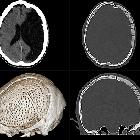
Kraniotomie

Ausgedehnte
Trepanation zur Druckentlastung nach Mediainfarkt mit Deckung durch Palacos.

Zustand nach
Operation / Erweiterungsplastik am Foramen magnum und Resektion der Kleinhirntonsillen bei asymmetrischer Chiari-1 Malformation. Man erkennt den erweiterten Liquorraum und Artefakte im okzipitalen Zugangsweg. T1 KM sagittal.


Computertomographie
sagittal im Hirnfenster und Knochenfenster sowie als Volumenrendering bei Zustand nach Erweiterungsplastik am Foramen magnum mit Resektion des hinteren Atlasbogens bei Chiari-1 Malformation.
A craniotomy is a surgical procedure where a piece of calvarial bone is removed to allow intracranial exposure. The bone flap is replaced at the end of the procedure, usually secured with microplates and screws. If the bone flap is not replaced it is either a craniectomy (bone removed) or cranioplasty (non-osseous surgical repair).
Classification
There are different craniotomy approaches depending on which part of the intracranial cavity needs to be accessed :
- frontal
- bifrontal
- parietal
- occipital
- pterional (i.e. frontosphenotemporal)
- subtemporal
- anterior parasagittal
- posterior parasagittal
- medial suboccipital
- lateral suboccipital
Complications
- infection including bone flap osteomyelitis, subdural empyema and cerebral abscess
- hemorrhage
- intracranial hemorrhage including remote cerebellar hemorrhage
- scalp/subcutaneous hematomas
- pseudomeningocoele
- tension pneumocephalus
Siehe auch:
und weiter:

 Assoziationen und Differentialdiagnosen zu Kraniotomie:
