Laminektomie

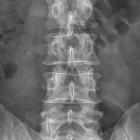
Röntgenbild
einer LWS mit Z. n. Laminektomie bei LWK 1. Der Dornfortsatz fehlt.

Bildbeschreibung:
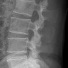
Hemilaminektomie Quelle: eigene Aufnahme Autor: User:Scuba-limp

Röntgenbild
der LWS nach Hemilaminektomie LWK 5 links. Der Dornfortsatz ist erhalten.

Z.n.
Laminektomie LWK 1 anamnestisch zur Tumorentfernung (z.B. Ependymom des Filum terminale?)

Z.n.
Laminektomie LWK 1 anamnestisch zur Tumorentfernung (z.B. Ependymom des Filum terminale?)

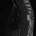
A rare cause
of acute paraplegia: granulocytic sarcoma. Sagittal spinal cord MRI T1: complete regression of compression process (the epidural mass was completely resected) with stigmata of laminectomy D6 (arrow)

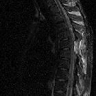
A rare cause
of acute paraplegia: granulocytic sarcoma. Sagittal spinal cord MRI T2: complete regression of compression process (the epidural mass was completely resected) with stigmata of laminectomy D6.
Laminectomy (whether unilateral or bilateral) refers to the surgical removal of the lamina of a vertebral body. By removing the lamina, we are able to decompress the spinal canal, and thus reduce the pressure on the spinal cord.
Spinal stenosis may be caused by:
The most commonly affected region is the lumbar spine.
A lumbar laminectomy is performed for patients with symptomatic, painful spinal stenosis, often occurring at multiple (> 3 vertebrae) levels of the spine.
The surgical procedure includes:
Siehe auch:
und weiter:

 Assoziationen und Differentialdiagnosen zu Laminektomie:

