leptomeningeale Tuberkulose

Extrapulmonary
tuberculosıs: an old but resurgent problem. Axial FLAIR (a), pre-contrast (b) axial T1 weighted, and post-contrast (c) axial T1 weighted MR images of a 21-year-old male. Axial FLAIR image shows high signal intensity caused by edema and inflammation in the left frontal lobe (star). In both anterior frontal regions (red arrows) and right parietal area (blue arrows) are seen subdural abscesses. In addition, MR images demonstrate meningeal enhancement and thickening (arrowheads)
Tuberculous
meningitis • Miliary tuberculosis involving the brain - Ganzer Fall bei Radiopaedia

Extrapulmonary
tuberculosıs: an old but resurgent problem. MR images of thoracic vertebrae of a 23-year-old female. Sagittal T2-weighted image reveals a heterogeneous signal increase in the spinal cord (red arrows) (a). Precontrast (b) and postcontrast (c) T1 weighted images of the same patient show linear (arrowheads) and nodular (curved arrow) dural enhancement and tuberculous abscess formations in the posterior epidural space

Magnetic
resonance imaging of sellar and juxtasellar abnormalities in the paediatric population: an imaging review. A 4-month-old baby boy with tuberculous meningitis. Axial (a, b) and coronal (c) post-contrast T1WI shows prominent extra-axial contrast-enhancement in the basal meninges, surrounding the brainstem, cerebellar sulci and Sylvian fissures. The leptomeningeal infection invades the left temporal lobe

Tuberculous
meningitis • Tuberculous leptomeningitis (gross pathology) - Ganzer Fall bei Radiopaedia

Tuberculous
meningitis • Tuberculous skull osteomyelitis with cerebral granulomas - Ganzer Fall bei Radiopaedia

Tuberculous
meningitis • Tuberculous meningitis with multiple infracts - Ganzer Fall bei Radiopaedia

Tuberculous
meningitis • Tuberculous meningitis - Ganzer Fall bei Radiopaedia

Tuberculous
meningitis • Tuberculous meningitis - Ganzer Fall bei Radiopaedia

Tuberculous
meningitis • Tuberculous meningitis with subarachnoid spinal involvement - Ganzer Fall bei Radiopaedia

Tuberculous
meningitis • Tuberculous meningitis - Ganzer Fall bei Radiopaedia

Leptomeningitis
• Tubercular meningitis with tuberculomas - Ganzer Fall bei Radiopaedia

Extrapulmonary
tuberculosis • Disseminated tuberculosis - Ganzer Fall bei Radiopaedia
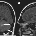
Tuberculous
meningitis: MRI findings.. Sagittal (A,B) FLAIR images showed areas with hyperintensity in the posterior fossa (left cerebellar peduncle and subarachnoid space) (arrows)

Tuberculous
meningitis: MRI findings.. Axial (A,B) FLAIR images showed areas with hyperintensity in the posterior fossa (left cerebellar peduncle and subarachnoid space) (arrows)

Tuberculous
meningitis: MRI findings.. T1 axial weighted images after gadolinium administration revealed nodular meningeal enhancement, around basal cisterns (arrows)


 Assoziationen und Differentialdiagnosen zu leptomeningeale Tuberkulose:


