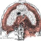
Ligamentum falciforme hepatis
The falciform ligament is a broad and thin peritoneal ligament. It is sickle-shaped and a remnant of the ventral mesentery of the fetus.
It is situated in an anteroposterior plane but lies obliquely so that one surface faces forward and is in contact with the peritoneum behind the right rectus abdominis and the diaphragm, while the other is directed backward and is in contact with the left lobe of the liver.
It contains between its layers a small but variable amount of fat and its free edge contains the obliterated umbilical vein (ligamentum teres) and if present, the falciform artery, and paraumbilical veins. The falciform ligament divides the left and right subphrenic compartments but may still allow passage of fluid from one to the other.
Arterial supply
Blood supply is very variable, and a separate hepatic falciform artery was only seen in 67% of cadavers in one study .
- left inferior phrenic artery and middle hepatic artery anastomose to form a single artery that then ramifies as 6-12 branches to supply the falciform ligament
Venous drainage
- left inferior phrenic vein drains the falciform ligament
History and etymology
The ligament derives its name from its shape, which is reminiscent of a sickle. Falciform is Latin for sickle-shaped. In the same way, the falx cerebri, is a sickle-shaped fold of dura separating the two cerebral hemispheres.
Siehe auch:
und weiter:

 Assoziationen und Differentialdiagnosen zu Ligamentum falciforme hepatis:
