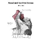
Maxillary bone


The maxillae (or maxillary bones) are a pair of symmetrical bones joined at the midline, which form the middle third of the face. Each maxilla forms the floor of the nasal cavity and parts of its lateral wall and roof, the roof of the oral cavity, contains the maxillary sinus, and contributes most of the inferior rim and floor of the orbit. Its alveolar process houses the teeth.
Gross anatomy
The body of the maxilla is roughly pyramidal and has four surfaces that surround the maxillary sinus, the largest paranasal sinus: anterior, infratemporal (posterior), orbital and nasal. It also has four processes: zygomatic, frontal, alveolar, and palatine. It articulates with the following bones: frontal, ethmoid, nasal, zygomatic, lacrimal, middle nasal concha, inferior nasal concha, palatine, and vomer.
Anterior surface
- has vertical protrusions overlying the roots of the teeth, with the canine eminence being the most prominent of these
- the incisive fossa runs medial to the eminence and the canine fossa is lateral to it
- above the canine fossa lies the infraorbital foramen, which transmits the infraorbital vessels and nerve
- above the infraorbital foramen lies the maxillary part of the infraorbital margin
- the anterior nasal spine is a vertical midline protuberance, with the nasal notch forming its deeply concave lateral border
Infratemporal surface
- forms the anterior wall of the infratemporal fossa
- on the inferior aspect of lateral margin, there may be a maxillary tuberosity, that appears after the appearance of the wisdom teeth
Orbital surface
- triangular in shape; forms most of orbital floor
- articulates with lacrimal bone, orbital plate of ethmoid, and orbital process of palatine bone
- posterior border forms most of anterior edge of inferior orbital fissure
- infraorbital groove passes centrally, transmits infraorbital vessels and nerve, and continues anteriorly as infraorbital canal, which opens into infraorbital foramen
- the canalis sinuosus, which transmits the anterior superior alveolar nerve and vessels, branches off of the infraorbital canal
Nasal surface
- maxillary ostium opens from maxillary sinus into hiatus semilunaris
- nasolacrimal groove is anterior to ostium; comprises two-thirds of the nasolacrimal canal circumference
- conchal crest articulates with inferior nasal concha
Zygomatic process
Pyramid-shaped projection at which anterior, infratemporal and orbital surfaces converge.
Frontal process
- located between the nasal and lacrimal bones
- contributes to the lacrimal fossa
- its medial surface is part of the lateral nasal wall
- closes anterior ethmoidal air cells
- articulates posteromedially with middle nasal concha, superiorly with nasal part of frontal bone, posterolateral with the lacrimal bone, and anteromedially with the nasal bone
Alveolar process
Contains eight sockets on each side for upper teeth; socket for canine tooth is deepest.
Palatine process
- horizontal; projects medially from lowest part of medial aspect of maxilla
- superior surface forms most of nasal floor
- inferior surface forms anterior three-fourths of hard palate
- contains two grooves posterolaterally that transmit the greater palatine vessels and nerves; additionally, many vascular foramina and depressions for palatine glands
- midline incisive fossa behind incisor teeth
- intermaxillary palatal suture runs posterior to the fossa
- two lateral incisive canals from nasal cavity open in incisive fossa and transmit terminations of greater palatine artery and nasopalatine nerve
See also
Siehe auch:
und weiter:

 Assoziationen und Differentialdiagnosen zu Maxilla:

