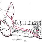
medial pterygoid muscle


The medial pterygoid muscle is one of the muscles of mastication.
Gross anatomy
The medial pterygoid muscle is a thick and square shaped muscle. It has two heads of origin. The deep head is the major component and is attached to the medial aspect of the lateral pterygoid plate of the sphenoid bone. The superficial head is attached to the maxillary tuberosity and the pyramidal process of the palatine bone.
The fibers of the medial pterygoid muscle run posterolaterally and insert to the medial surface of the mandibular ramus and angle.
Actions
The medial pterygoid acts together with masseter to elevate the mandible.
Acting together with the lateral pterygoid muscles, they protrude the mandible which is important in the grinding movement of mastication.
Blood supply
The arterial supply to medial pterygoid muscle is from the pterygoid branches of the maxillary artery.
Innervation
Medial pterygoid nerve of the mandibular division of the trigeminal nerve (CN V3).
Variations
The muscles of mastication are all derived from the 1st pharyngeal arch, so fusions between these muscles may be observed.
The medial pterygoid may send a fascicle to the masseter which gives rise to the styloglossus.
Siehe auch:

 Assoziationen und Differentialdiagnosen zu Musculus pterygoideus medialis:
