mesenteric cyst

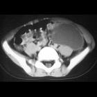
Newborn with
palpable lower abdominal massAxial CT with contrast of the abdomen shows a large simple cystic mass in the left side of the abdomen.The diagnosis was mesenteric cyst.

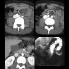
Teenager with
abdominal pain. Two contiguous axial images from a CT with contrast of the abdomen show a whirlpool sign of swirling of the mesenteric vessels (above left) around a round low density structure just to the left of the spine (above right). Another axial image from the same exam (below left) shows a normal relationship of the superior mesenteric vein to the right of the superior mesenteric artery although both appear to be shifted to the left. AP image from a subsequent upper GI (below right) shows normal position of the ligament of Treitz in the left upper quadrant.The diagnosis was small bowel volvulus around a mesenteric cyst. There was no evidence of malrotation or midgut volvulus.

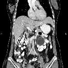
Teenager with
a cystic abdominal mass. Surgical image shows a large thin-walled cystic structure arising from the mesentery of the small bowel.The diagnosis was mesenteric cyst.

Mesenteric
cyst • Mesenteric cystic lymphangioma - Ganzer Fall bei Radiopaedia

Mesenteric
cyst • Mesenteric cyst - Ganzer Fall bei Radiopaedia

Mesenteric
cyst • Mesenteric cyst - Ganzer Fall bei Radiopaedia

Mesenteric
cyst • Mesenteric cystic lymphangioma - Ganzer Fall bei Radiopaedia

Mesenteric
cyst • Cystic lymphangioma - Ganzer Fall bei Radiopaedia

Mesenteric
cyst • Mesenteric cyst - Ganzer Fall bei Radiopaedia

Mesenteric
cyst • Calcified cystic mesenteric lymphangioma - Ganzer Fall bei Radiopaedia

Mesenteric
cyst • Mesenteric lymphangioma - Ganzer Fall bei Radiopaedia

Mesenteric
cyst • Giant mesenteric cyst - Ganzer Fall bei Radiopaedia

Mesenteric
cyst • Mesenteric cyst - Ganzer Fall bei Radiopaedia

Mesenteric
cyst • Mesenteric cyst - Ganzer Fall bei Radiopaedia

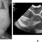
Teenager with
chronic abdominal pain. Image from a small bowel follow through exam (above) shows the small bowel to be entirely opacified and to be elevated out of the pelvis with the exception of a single loop in the middle of the pelvis. Axial CT without intravenous and with oral contrast of the abdomen (below) shows a large low density multiseptated mass in the pelvis.The diagnosis was mesenteric cyst.
Mesenteric cysts are a very rare cause of abdominal pain and have a wide range of underlying causes.
Epidemiology
Mesenteric cysts are rare, with a reported incidence of 0.5-1 per 100,000 admissions .
Clinical presentation
Patients usually present with abdominal pain and/or mass, although the cysts can be asymptomatic, incidental finding .
Pathology
Etiology
- lymphatic: simple lymphatic cyst and lymphangioma
- mesothelial: simple mesothelial cyst, benign cystic mesothelioma, and malignant cystic mesothelioma
- enteric: enteric cyst and enteric duplication cyst
- urogenital
- mature cystic teratoma (dermoid cysts)
- non-pancreatic pseudocysts (infectious and traumatic cysts)
Radiographic features
Mesenteric cysts can occur anywhere in the mesentery, from the duodenum to the rectum, and may extend into the retroperitoneum. Mesenteric cysts are cystic mesenteric lesions that can be further characterized by the wall thickness (thin or thick-walled) and their loculation (unilocular or multilocular).
Differential diagnosis
The main differential diagnosis is:
- pancreatic pseudocyst: usually not only abuts but involves the pancreas directly, and communicates with the main pancreatic duct
- ovarian cyst
- meconium pseudocyst
- urachal cyst
- hydrometrocolpos
- peritoneal hydatidosis
Siehe auch:
- Duplikationszysten gastrointestinal
- zystisches Lymphangiom des Mesenterium
- fetale enterale Duplikaturen
und weiter:

 Assoziationen und Differentialdiagnosen zu mesenteric cyst:


