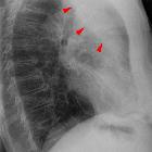
middle lobe collapse


Right middle lobe collapse (or simply termed middle lobe collapse) has distinctive features, but can be subtle on frontal chest radiographs.
For a general discussion please refer to the article on lobar collapse.
It is important to note that of all the lobes, the right middle lobe is the most likely to be chronically collapsed. When this is the case it is referred to as right middle lobe syndrome .
Radiographic features
Plain radiograph
On frontal radiographs the findings are subtle compared to the lateral projection and include :
- right mid to lower zone air space opacification (which can be subtle)
- the normal horizontal fissure is no longer visible (as it rotates inferiorly rendering it non-tangental to the x-ray beam
- a lordotic view may help identify the displaced fissure
- obscuration of the right heart border
- increased opacity adjacent to the right heart border requires a degree of consolidation as well as atelectasis
Non-specific signs indicating right sided atelectasis may also be present although due to the small size of the right middle lobe they may well be subtle or absent :
- elevation of the hemidiaphragm
- crowding of the right sided ribs
- shift of the mediastinum to the right
- displacement of the right hilum
On lateral projection, right middle lobe collapse is usually relatively easy to identify, appearing as a triangular opacity in the anterior aspect of the chest overlying the cardiac shadow, with its apex at the right hilum. The horizontal fissure is displaced inferiorly and the inferior part of the oblique fissure is displaced and bowed anterosuperiorly.
Presence of tubular lucencies in the collapsed right middle lobe should suggest that the collapse is chronic (right middle lobe syndrome), with associated bronchiectasis .
If there is an obstructing lesion in the bronchus intermedius, there will be signs of both RML and RLL.
CT
- triangular opacification in axial images abutting the right heart border, thinner at the hilum, and the apex pointing laterally
- horizontal fissure rotates anteromedially
- oblique fissure bows anteriorly
- RUL rotates anterolaterally and RLL rotates posterolaterally and meet lateral to the collapsed RML
Differential diagnosis
Frontal projection
On frontal (PA or AP) projection, right middle lobe collapse should be distinguished from:
- right middle lobe consolidation: absence of signs of volume loss
- pectus excavatum: downward sloping ribs and shift of the heart away from the right are clues; lateral projections make the distinction easy
Lateral projection
On the lateral projection, right middle lobe collapse should be distinguished from:
- fluid within the oblique fissure (pseudotumor): horizontal fissure should be visible as separate to the opacity
- fat within the oblique fissure
- consolidation of the right middle lobe
Related articles
Siehe auch:
- Atelektase
- right middle lobe consolidation
- Pectus excavatum
- left lower lobe collapse
- Lady Windermere Syndrom
- right upper lobe collapse
- left upper lobe collapse
- Mittellappenpneumonie
- Unterlappenatelektase rechts
- lobar collapse
- Normale Herzkonfiguration im Röntgen-Thorax
- Mittellappen-Syndrom
und weiter:

 Assoziationen und Differentialdiagnosen zu Right middle lobe atelectasis:


