Milztorsion


Splenic volvulus (rare plural: volvuli) may be seen as a complication of a wandering spleen due to weakness of the splenic ligaments .
Clinical presentation
- abdominal pain: mild to severe in intensity which depends on the degree of torsion
- abdominal mass
- abdominal discomfort
- acute abdomen
Pathology
Laxity of splenic ligaments may lead to a wandering spleen . The spleen is identified anywhere in the abdomen or in the pelvis that ultimately results in splenic torsion that will lead to congestion, torsion, infarction or in severe cases splenic rupture .
The spleen wanders due to increased mobility-related to ligamentous laxity of its peritoneal attachments. Various factors of ligamentous laxity which encompass congenital and acquired etiology. In the congenital form, there is the failure of development of the normal splenic suspensory ligaments, including the lienorenal and gastrosplenic ligaments. The acquired form can happen in situations that weaken the ligaments, such as pregnancy (with subsequent high estrogen level) or trauma.
Radiographic features
Diagnosis is primarily done with Doppler ultrasound and CT .
Ultrasound
Ultrasonography reveals a round, hypoechoic, solid-looking mass in an ectopic or orthotopic location .
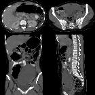
CT
CT scan is the gold standard test in cases of splenic volvulus .
- ectopically located or malpositioned spleen
- splenic vascular pedicle demonstrates a whirlpool configuration
- contrast-enhanced CT demonstrates a low-density spleen with peripheral enhancement or no enhancement
Treatment and prognosis
Open or laparoscopic splenopexy is a spleen-saving technique that can be performed in cases of lack of infarction. Surgical intervention is mandatory in most of the cases .
See also
Siehe auch:


 Assoziationen und Differentialdiagnosen zu Milztorsion:
