Morbus Sudeck


Complex regional pain syndrome (CRPS), also known as Sudeck atrophy, is a condition which can affect the extremities in a wide clinical spectrum. CRPS is principally a clinical diagnosis seen more commonly in females than males with a mean age of presentation of 50 to 70 years . No one imaging study is sensitive or specific to rule in or rule out the syndrome.
Terminology
Two types of CRPS have been described :
- type 1: no underlying single nerve lesion (formerly known as reflex sympathetic dystrophy)
- type 2: underlying nerve lesion identified (formerly known as causalgia)
Clinical presentation
Patients present after an initiating event (see causes below) with symptoms of more than 6 months duration such as edema, changes in skin blood flow, abnormal motor activity, allodynia or hyperalgesia . Symptoms are often out of proportion to the initiating event and not limited to a single peripheral nerve .
Pathology
Etiology
- trauma: often minor
- surgery
- idiopathic: immobilization
- unknown in many cases
- CNS disorders
- myocardial infarction
Location
Occurs in hands and feet distal to the injury.
Radiographic features
Plain radiograph
- severe patchy osteopenia, particularly in the periarticular region
- soft tissue swelling, with eventual soft tissue atrophy
- subperiosteal bone resorption
- preservation of joint space
It is important to differentiate this from disuse osteopenia since the clinician could initiate aggressive physical therapy for the latter.
MRI
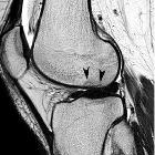
- patchy bone marrow edema signal (particularly subcortical), although bone marrow signal may be normal in some cases
- soft tissue edema and enhancement
- skin thickening
- joint effusion
- synovial hypertrophy
- muscle atrophy in later stages
Nuclear medicine
- presence of CRPS can be evaluated with a 3-phase bone scan
- the classical presentation is increased biphosphonate tracer uptake in the affected limb on all three phases (seen less than 50% of the time, but has the highest diagnostic accuracy)
- diffusely increased juxta-articular activity around all joints of the affected hand or foot on delayed images is the most sensitive indicator (infection and arthritis are potential false-positive findings)
Treatment and prognosis
In most cases, a multidisciplinary approach is required whereby a combination of various treatments may be employed, such as physical therapy, systemic or regional medications, sympathectomy or spinal cord stimulation, and psychotherapy. Interventional radiology can offer pain relief by peripheral nerve block procedures.
Siehe auch:
und weiter:

 Assoziationen und Differentialdiagnosen zu Morbus Sudeck:


