morton neuroma


Morton neuromas, also known as interdigital or intermetatarsal neuromas, are focal areas of symptomatic perineural fibrosis around a plantar digital nerve of the foot. The condition is thought to be due to chronic entrapment of the nerve by the intermetatarsal ligament.
Terminology
The term neuroma is a misnomer because the abnormality is non-neoplastic and does not represent a true neuroma. It may more correctly be known as Morton metatarsalgia.
Epidemiology
It most often occurs in middle-aged individuals and is many times more common in women than men. Approximately 30% of asymptomatic middle-aged persons have the radiologic-pathologic findings of a Morton neuroma.
Clinical presentation
Patients typically present with forefoot pain which radiates from midfoot to toes . Symptoms are often progressive and worsened by activity. The Mulder sign is a physical sign associated with Morton neuroma, which may be elicited while the patient is in a supine position. The pain associated with the neuroma, as well as a click, can be reproduced by squeezing the two metatarsal heads together with one hand, while concomitantly putting pressure on the interdigital space with the other hand.
A number of other clinical tests for Morton neuroma have been described, all of which have high specificity and aim to either reproduce symptoms of pain or paresthesia. These include :
- thumb-index finger squeeze: squeeze the symptomatic intermetatarsal space between the index (dorsal) and thumb (plantar) surfaces
- foot squeeze test: compression of metatarsal heads between fingers and thumb
- plantar and dorsal percussion tests: percussion of the dorsal and plantar intermetatarsal spaces with a finger
- light touch sensory test: stroking the tip of the affected toe produces sensation different to the adjacent toes
Pathology
It is characterized by neural degeneration with epineural and endoneural vascular hyalinisation, and perineural fibrosis around a plantar digital nerve .
Location
The 3 web-space (between 3 and 4metatarsal heads) is the most commonly affected site. The 2 web-space is less often involved while the remaining web-spaces are rarely involved. 10% of lesions are bilateral.
Occasionally specific terms are used when occurring certain spaces
- 1 intermetatarsal space: Heuter neuroma
- 4intermetatarsal space: Iselin neuroma
Radiographic features
Symptomatic lesions tend to be slightly larger (mean 5.3 mm vs. 4.1 mm in one large series ). Lesions >5 mm are very likely to be symptomatic.
Ultrasound
Typically seen as a round to ovoid, well-defined, hypoechoic lesion in the intermetatarsal space proximal to the metatarsal head . A Morton neuroma is not compressible. A small proportion can have mixed echotexture . A sonographic Mulder sign may be elicited with the probe .
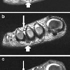
MRI
Dumbbell/ovoid-shaped lesion at a similar position to that described on ultrasound :
- T1: typically low-to-iso signal
- T2: typically low signal but can sometimes be intermediate in signal
- T1 C+ (Gd): variable enhancement
Treatment and prognosis
Ultrasound-guided interdigital injection of steroid and local anesthetic has been demonstrated to have a relatively high success rate .
Surgical excision can also be performed, also with a relatively high success rate (~80% ).
History and etymology
It is named after Thomas George Morton (1835-1903), an American surgeon, who described a case series in 1876 . However, it was first described by Civinini in 1835 .
Differential diagnosis
Other causes of metatarsalgia:
- intermetatarsal bursa/bursitis
- extruding out in between the metatarsal bones on the plantar aspect of the foot
- compressible
- metatarsophalangeal joint synovitis/capsulitis
- changes secondary to a plantar plate tear/disruption
- soft tissues tumors, e.g. schwannoma
- metatarsal head insufficiency fracture / Freiberg disease
- tenosynovial giant cell tumor
Siehe auch:
und weiter:


 Assoziationen und Differentialdiagnosen zu Morton-Neuralgie:
