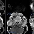
mukoepidermoides Karzinom der Parotis

Imaging of
parotid anomalies in infants and children. Intermediate-grade mucoepidermoid carcinoma: poorly delineated, heterogeneous tumor containing small necrotic areas [white arrow] and strongly enhanced, on ultrasonography (a and b), axial T2-weighted image (c) and T1-weighted image, after intravenous injection of gadolinium chelate (d). Time intensity curve with a signal growing rapidly in the first phase and a low wash out (< 30%): type C or plateau pattern (according to Yabuuchi), suggestive of malignancy (e). Conversely, time intensity curve of type B (orange line), with a high wash out (> 30%), in a case of parotid benign lymphadenopathy (f)
mukoepidermoides Karzinom der Parotis
Siehe auch:

 Assoziationen und Differentialdiagnosen zu mukoepidermoides Karzinom der Parotis:
mukoepidermoides
Karzinom der Speicheldrüsen


