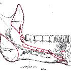
Musculus mylohyoideus


An anatomical
illustration from the 1909 American edition of Sobotta"s Atlas and Text-book of Human Anatomy with English terminology

An anatomical
illustration from the 1909 American edition of Sobotta"s Atlas and Text-book of Human Anatomy with English terminology


Imaging of
the sublingual and submandibular spaces. Anatomy of the submandibular and sublingual spaces in the coronal plane: picture illustration and T2-weighted MR image
The mylohyoid muscles form a paired muscular sling that forms part of the floor of the mouth. It also separates the sublingual space (and oral cavity) from the submandibular space.
Summary
- origin: mylohyoid line/ridge on the medial surface of the mandible
- insertion: midline raphe that extends from the mandibular symphysis to the hyoid bone
Arterial supply
- sublingual branch of the lingual artery
- mylohyoid branch of the inferior alveolar artery
- submental branch of the facial artery
Innervation
- mylohyoid nerve, a branch of the inferior alveolar nerve
Action
- elevates the floor of the mouth (e.g. in swallowing or protruding the tongue)
Variant anatomy
- mylohyoid boutonniere: defect in the midportion of the mylohyoid muscle through which sublingual glands, blood vessels, fat, etc. can herniate
Siehe auch:
und weiter:

 Assoziationen und Differentialdiagnosen zu Musculus mylohyoideus:

