occult fracture


Im
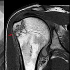
Röntgenbild nicht erkennbare Fraktur des Oberarmkopfes. In der Magnetresonanztomographie zeigt sich das Ödem im Knochen je nach Sequenz dunkel (T1 Mitte) oder hell (PDW SPIR rechts).

Fat pad sign:
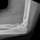
Ventral fat pad bowed and dorsal fat pat visible. Non displaced fracture of the radius head.

Primär
(linkes Bild) okkulte Fraktur der Fibula. Nach ca. 3 Wochen bei anhaltenden Beschwerden zeigt sich in der Kontrolle (rechts) die Fraktur jetzt mit leichtem Versatz. Beachte auch die Schwellung.

Liphämarthros
Schulter bei fast okkulter Fraktur: Die Röntgenaufnahme zeigt oberhalb des tief stehenden Humeruskopfes einen waagerechten Spiegel. Fraktur nur am medialen Rand erahnbar. In der 2 Tage darauf angefertigten MRT bestätigt sich die kaum verschobene Fraktur. Das intraartikuläre Fett ist schon nicht mehr nachweisbar.

School ager
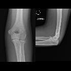
with elbow pain after falling on their arm. AP (left) and lateral (right) radiographs of the elbow show elevation of the anterior fat pad on the lateral view. No fracture line is seen.The diagnosis was occult elbow fracture.

Occult
fracture • Subtle distal radius fracture - Ganzer Fall bei Radiopaedia

Occult
fracture • Radial head occult fracture - Ganzer Fall bei Radiopaedia
Occult fractures are those that are not visible on imaging, most commonly plain radiographs and sometimes CT, either due to lack of displacement or limitations of the imaging study. There may be clinical signs of a fracture without one actually being seen. MRI or nuclear medicine studies are sometimes required to make the diagnosis.
Technically any fracture may be occult, but classic examples include:
- distal radius fracture (pronator quadratus fat pad sign may be positive)
- neck of femur fracture
- radial head fracture (sail sign may be positive)
- scaphoid fracture
- supracondylar fracture in children (loss of alignment may be the only sign)
Siehe auch:
und weiter:

 Assoziationen und Differentialdiagnosen zu Okkulte Fraktur:


