Os cuneiforme mediale

The medial cuneiform is one of the tarsal bones located between the navicular and base of first metatarsal, medial to the intermediate cuneiform bone.
Gross anatomy
Osteology
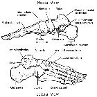
The medial cuneiform is one of the cuneiforms, it is the most medial in the distal row of tarsal bones.
It has a narrow dorsal surface and a flat plantar surface which receives a slip from the tibialis posterior tendon. The distal surface is reniform, congruent with the articulating base of first metatarsal and proximal surface has a pyriform facet for the navicular. The medial surface is subcutaneous, convex vertically and exhibits an impression for the tibialis anterior. The lateral surface shows dorsal facets for the articulations with intermediate cuneiforms and second metatarsal base. The distal plantar aspect of the lateral surface demonstrates a groove for the peroneus longus tendon.
Articulations
As described above, the medial cuneiform exhibits various facets separated by bony ridges, articulating with the first and second metatarsal bases, the intermediate cuneiform and navicular bones.
Attachments
Musculotendinous
These are mainly along the plantar aspect:
- tibialis posterior tendon insertion at proximal margin
- third plantar interosseous muscle origin along lateral margin
- peroneus longus tendon part insertion at plantar aspect of the lateral margin
- tibialis anterior tendon insertion at medial margin
Ligamentous
The plantar aspect of the lateral surface shows ligamentous attachments to the first, second and third metatarsal bases via plantar tarsometatarsal ligaments and receives a slip from the plantar cuneonavicular ligament.
Proximally it is attached to the intermediate cuneiform through interosseous intercuneiform ligament.
Arterial supply
This bone receives medial, lateral and dorsal surface arterial branches from the dorsal arterial network .
Venous drainage
The draining veins of the medial cuneiform correspond to the arterial supply.
Innervation
It is supplied by the deep peroneal and medial plantar nerves.
Variant anatomy
- bipartite medial cuneiform
- sesamoid bones associated with medial cuneiform are os intercuneiforme and pars peronea metatarsalis
Development
Ossification
This bone usually has one ossific center but may have two centers in some cases. The ossific center appears at two years of age.
Siehe auch:
und weiter:


 Assoziationen und Differentialdiagnosen zu Os cuneiforme mediale: