bipartite medial cuneiform


A bipartite medial cuneiform is an anatomical variant where there are two ossification centers involving the medial cuneiform. In many cases, the overall shape of the medial cuneiform is conserved, although the size of the two combined bones is larger than that of a normal medial cuneiform.
Epidemiology
The estimated incidence is thought to be ~1% (range 0.3-2.4%) of the general population with the majority (~85%) reported in males .
Associations
Some patients may have additional associations which include:
- os intermetatarseum
- accessory navicular bone
- spinal segmentation anomalies
- lunotriquetral coalition
- an accessory temporal bone suture
- bipartite temporal bone
Clinical presentation
Most patients are asymptomatic although occasionally some patients can present with pain.
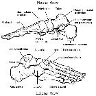
Gross anatomy
Bipartite medial cuneiforms are divided by an oblique/horizontal synchondrosis and/or syndesmosis into a smaller dorsal ossicle and larger plantar ossicle .
Radiographic features
Plain radiograph
A lateral foot radiograph (30° angled) may give an "E" sign with the two bones most often dorsal and plantar viewed end on.
MRI
May also give an "E" sign when viewed on sagittal images. MRI also allows direct visualization of anatomy as well as an appreciation of any signal abnormality along the synchondrosis.
Treatment and prognosis
Symptomatic cases may benefit from intervention ranging from various nonoperative measures to surgical excision.
See also
Siehe auch:


 Assoziationen und Differentialdiagnosen zu Os cuneiforme mediale bipartitium: