Ossifikationen der Falx


In discussing mineralization of the falx cerebri, many radiology textbooks use the term falx calcification and make no mention of falx ossification.
Epidemiology
Ossification of dural folds is relatively unusual; one study suggested a prevalence of falx ossification of 0.7% . Even though, ossification of an isolated site of the falx cerebri is very rare in medical literature .
None of the available literature had mentioned whether the gender or ethnicity plays any role in falx ossification.
Associations
Incidence of falcine ossification has been reported in certain medical disorders such as :
- endocrine disorders (e.g. hyperparathyroidism)
- vitamin D intoxication
- chronic renal failure
- basal cell nevus syndrome
- Maroteaux type brachyolmia
- hypertelorism
- pseudoxanthoma elasticum
Pathology
Since the falx cerebri is derived from embryonic mesenchymal cells, occasional ossification might be seen due to friction, hemorrhage, or trauma, which results in some osteogenic changes leading to the formation of membranous bone.
Radiographic features
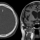
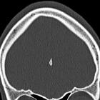
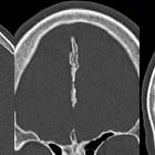
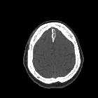
CT
CT scan can clearly distinguish an isolated ossification of the falx from ossification that has spread to the falx from the frontal bone .
MRI
On magnetic resonance images, falx ossification exhibits a typical appearance consisting of a central marrow-containing portion with signal intensity similar to fat (bright on T1), surrounded by low signal intensity representing cortical bone .
Differential diagnosis
There is no difficulty identifying the ossification process on CT images, nevertheless on MRI images some differentials should be considered:
- fatty falx cerebri
- falx cerebri calcification
- hemorrhage (in a trauma context)
See also
Siehe auch:
und weiter:


 Assoziationen und Differentialdiagnosen zu Ossifikationen der Falx:

