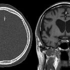
Falxlipom


 nicht verwechseln mit: Balkenlipom
A fatty falx cerebri is a benign entity in which there is fat within the extradural neural axis compartment located between the two visceral layers of the falx.
Epidemiology
According to one study, it is a common finding seen in approximately 7.3% of patients . This can be more common in older patients .
Clinical presentation
Patients are usually asymptomatic and found incidentally.
Radiographic features
The characteristic finding on both CT and MRI is fat within the falx cerebri.
CT
On CT, there is midline homogenous fat attenuation (hence negative CT attenuation values) in the falx cerebri.
MRI
MRI with and without fat saturation are able to make the diagnosis easily.
Signal characteristics are that of fat:
- T1: high signal intensity
- T2: high signal intensity
- T1 C+ (Gd): no enhancement
- Fat saturated sequences: low signal
Treatment and prognosis
No treatment is recommended as this is an asymptomatic and incidental finding.
Differential diagnosis
A fatty falx is an incidental finding and should not be mistaken for:
- ruptured intracranial dermoid: multiple droplets of fat in the subarachnoid space
- intracranial lipoma: frequently inter-hemispheric and associated agenesis of the corpus callosum
- pericallosal lipoma
- osseous metaplasia of the falx cerebri: fat within the medullary space of an ossified falx (also common)
- pneumocephalus: ROI should be no lower than -120HU (beware volume averaging)
See also
Siehe auch:
- Falxmeningeom
- Balkenlipom
- Pneumocephalus
- intrakranielle Lipome
- Ossifikationen der Falx
- intrakranielles Fett
- Dysgenesie des Corpus callosum
- rupturierte intrakranielle Dermoidzyste
- Lipome in den intrakranialen venösen Sinus
und weiter:

 Assoziationen und Differentialdiagnosen zu Falxlipom:


