osteoradionecrosis


Osteoradionecrosis (ORN) refers to a severe delayed radiation-induced injury and is characterized by bone tissue necrosis and failure in healing. There is some overlap with the term radiation osteitis.
Pathology
Microvascular damage is thought to result in altered blood supply to the bone. This limits the ability of the tissues to repair normal wear and tear which, in turn, eventually results in the tissue breakdown seen on imaging.
Usually, large radiation doses are required for osteoradionecrosis to occur (e.g. <3000-5000 cGy) .
Location
While it can involve any bone within the irradiated field, there are specific sites in which osteoradionecrosis is more commonly seen. These include:
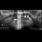
- mandible: mandibular osteoradionecrosis: this site is particularly prone due to its superficial location and high doses of radiation required to radically treat naso-oropharyngeal tumors
- chest wall-shoulder-humerus-scapula
- spine
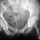
- bony pelvis
Radiographic features
While there are general features, radiographic features can somewhat vary with the site of involvement.
Plain radiograph
With mandibular osteoradionecrosis, there can be ill-defined cortical destruction without sequestration. In osteoradionecrosis of the ribs, clavicle, scapula, and humerus, radiography may demonstrate :
- osteopenia: typically occurs after ~1 year postirradiation
- disorganization and coarsening of trabecular architecture
- cortical irregularity
- heterogenous bone density
CT
With mandibular osteoradionecrosis, CT may additionally show cortical interruptions and loss of spongiosa trabeculation. In other sites, CT may show the presence of subtle fractures, alterations in bone architecture and dystrophic soft-tissue calcification.
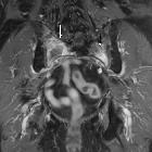
MRI
On MRI, they can be the development of new heterogeneous signal within the marrow of an irradiated area (intermediate or low T1 signal, intermediate or high T2 signal). Osteoradionecrosis with or without osteomyelitis can be extremely difficult to differentiate from a recurrent tumor. Adjacent muscles may appear edematous and show intense enhancement, which can be difficult to differentiate from recurrent tumor if bone changes are not visible on CT .
In osteoradionecrosis of the spine, hematopoietic cellular elements of the spinal marrow can also be replaced with fat, which then has
- T1: high signal intensity
- T2: intermediate signal intensity
Treatment and prognosis
It either stabilizes or gradually worsens, which then becomes notoriously difficult to manage.
Differential diagnosis
To give a meaningful differentiation, location and imaging modality needs to be taken into account. General differential consideration includes:
- original tumor recurrence
- radiation-induced secondary tumor e.g. sarcoma
- complicating infection: osteomyelitis (can also be an association)
See also
Siehe auch:
und weiter:


 Assoziationen und Differentialdiagnosen zu Osteoradionekrose:
