zerebrale Radionekrose


Cerebral radiation necrosis refers to necrotic degradation of brain tissue following intracranial or regional radiation either delivered for the treatment of intracranial pathology (e.g. astrocytoma, cerebral arteriovenous malformation) or as a result of irradiation of head and neck tumors (e.g. nasopharyngeal carcinoma).
Terminology
Although post-radiation treatment effects include pseudoprogression, which seen relatively close to radiation particularly in the setting of concurrent chemotherapy for glioblastoma (Stupp protocol). This article focuses on more delayed effect, generally termed radiation necrosis, which appears months to several years after radiation therapy and involves a space-occupying necrotic lesion with mass effect and neurological dysfunction.
Pathology
There are numerous potential pathways to radiation necrosis which include:
- vascular injury
- acutely endothelial damage can lead to vasogenic edema
- chronically fibrosis, hyalinization and stenosis can occur with eventual thrombosis and infarction
- vascular ectasia and telangiectasia are also seen frequently, with capillary telangiectasias and cavernous malformations common findings post whole brain irradiation.
- oligodendrocytes and white matter damage
- oligodendrocytes are sensitive to radiation
- loss of white matter accounts for the majority of volume loss
- effects on the fibrinolytic enzyme system
- increase in urokinase plasminogen activator and a simultaneous decrease in tissue plasminogen activator may contribute to cytotoxic edema and tissue necrosis
- immune mechanisms
Radiographic features
MRI
- T2/FLAIR: white matter high signal
- edema and mass effect early
- loss of volume later
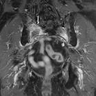
- T1 C+ (Gd)
- white (more common) or grey matter
- single or multiple
- nodular or curvilinear
- "soap-bubble", “cut green pepper” or "Swiss-cheese" enhancement
- occasionally can be ring enhancing (see MAGIC DR mnemonic)
- MR spectroscopy: typically low choline, creatine, and NAA
- MR perfusion: areas of enhancement and high T2/FLAIR don't show increased rCBV in radiation necrosis or pseudoprogression and could be helpful in distinguishing them from residual lesion or recurrence
FDG-PET
- radiation necrosis is usually hypometabolic whereas tumor is hypermetabolic
Practical points
It has been suggested that involvement of the corpus callosum with the crossing of the midline and multiple lesions or subependymal spread would favor a recurrent tumor over radiation necrosis , however, conventional imaging can be misleading, and no individual feature is reliable.
Siehe auch:
- Corpus callosum
- zerebrale arteriovenöse Malformation
- Astrozytom
- Nasopharynxkarzinom
- zerebrale Läsionen mit ringförmiger Kontrastmittelanreicherung
- Osteoradionekrose
- Strahlennekrose
- MAGIC DR
und weiter:


 Assoziationen und Differentialdiagnosen zu zerebrale Radionekrose: