ovarian borderline serous cystadenoma


Borderline ovarian serous cystadenomas lie in the intermediate range in the spectrum of ovarian serous tumors and represent approximately 15% of all serous tumors.
Epidemiology
They present at a younger age group than the more malignant serous cystadenocarcinomas with a peak age of presentation of ~45 years of age .
Clinical presentation
The tumors are often clinically silent until they achieve an advanced size or stage. The most frequent initial manifestations were abdominal pain, increasing abdominal girth or distension, or as an abdominal mass .
Pathology
Borderline tumors fall under ovarian epithelial tumors. They tend to develop in an exophytic growth pattern, on the surface of the ovary, without invading the underlying stroma. Papillary projections are characteristic and may be more of a feature with borderline than malignant serous cystadenocarcinoma of the ovary.
A unique feature of borderline tumors is the non-invasive behavior of extra-ovarian tumor implants in the advanced stages of the disease . Implants can occur in the contralateral ovary, omentum, and peritoneal surface in the advanced stages, although they behave in a benign fashion and remain located on the surface of the underlying tissues.
Markers
Serum CA-125 level is typically mildly elevated.
Radiographic features
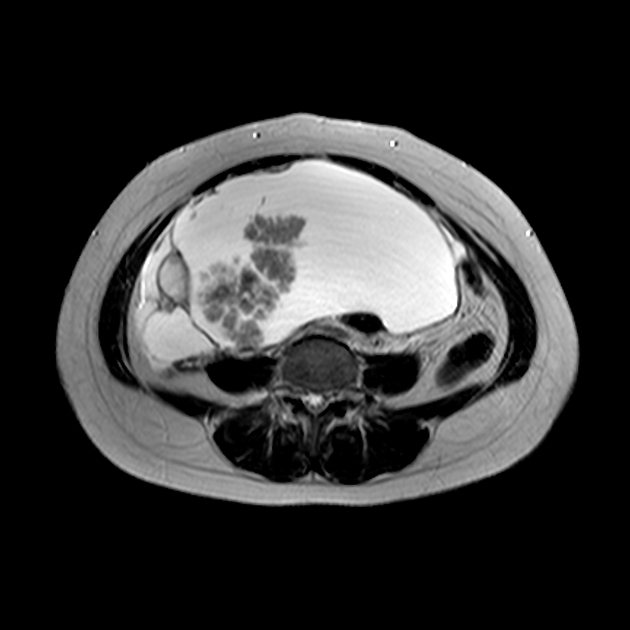
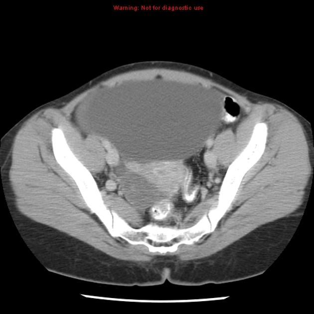
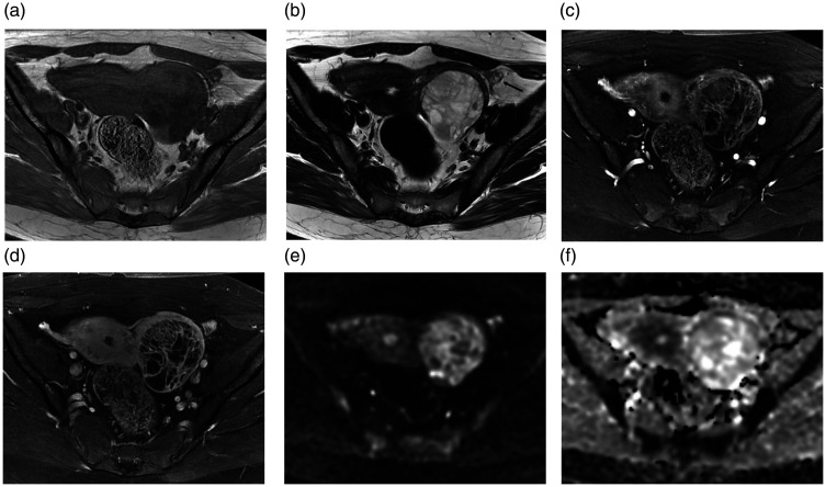
Typically seen as bilateral adnexal masses with profuse papillary projections. Bilaterality occurs more frequently than with benign ovarian serous cystadenomas .
Serous borderline tumors may display aggressive behavior, and occasionally present with peritoneal or nodal metastases.
Ultrasound
The rate of detection of intratumoral blood flow on Doppler ultrasound can be very similar to more malignant neoplasms .
Treatment and prognosis
Post-surgical prognosis is better than for ovarian cystadenocarcinoma, even in the presence of transovarian spread .
Staging
Borderline tumors are staged using the same ovarian cancer staging as malignant ovarian neoplasms.
History and etymology
They were first described in 1929 and were designated for separate classification in the early 1970s by the World Health Organization .
Siehe auch:
- Neoplasien des Ovars
- ovarian serous cystadenocarcinoma
- Ovarialkarzinom Staging
- ovarian serous tumours
- seröses Zystadenom des Ovars
- serous cystadenocarcinomas
- Endosalpingiose
und weiter:

 Assoziationen und Differentialdiagnosen zu seröses Boderline-Zystadenom des Ovars:


