ovarian serous cystadenoma


Ovarian serous cystadenomas are a type of benign ovarian epithelial tumor at the benign end of the spectrum of ovarian serous tumors.
Terminology
Serous ovarian tumors are traditionally described with a "cyst-" prefix because of their primarily cystic composition, e.g. cystadenoma, cystadenocarcinoma.
Epidemiology
Serous cystadenomas account for ~60% of ovarian serous tumors . They are the commonest type of ovarian epithelial neoplasm. The peak incidence is at the 4 to 5 decades of life.
Clinical presentation
Generally asymptomatic. If symptoms are present, they are usually related to mass effect with displacement of adjacent structures, e.g. loops of bowel, adnexal torsion.
Pathology
Etiology
Thought to largely derive from ovarian epithelial inclusions, which itself is derived from fallopian tube epithelium.
Location
They can be bilateral in ~15% of cases.
Macroscopic appearance
Serous cystadenomas are usually composed of unilocular (or at times multilocular) cysts filled with clear watery fluid. The cysts measure 10 cm in average diameter but may be extremely large. The lining of the cyst is flat or may contain small papillary projections.
Microscopic appearance
Composed of a solitary layer of benign epithelium which resembles fallopian tube mucosa and may form papillary structures. As with other serous tumors, psammomatous calcification can be a feature.
Radiographic features
Ultrasound
- usually seen as a unilocular cystic/anechoic adnexal lesion
- papillary projections are absent
- if there is any wall irregularity, it is thin, with an acute angle with the cyst wall and has a regular surface
- some lesions may contain sonographically detectable septations
- no flow is detected on color doppler
- higher resistive and pulsatility indices on pulsed doppler when compared to malignant neoplasms
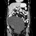
CT
Often seen as a unilocular (typically) or multilocular cystic mass with homogeneous CT attenuation, with a thin regular wall or septum, and usually no endocystic or exocystic vegetation . Cysts can be quite large in size and have the potential to be seen filling most of the lower pelvis with extension into the upper abdomen.
MRI
The typical MR imaging appearance of serous cystadenoma is a unilocular thin-walled adnexal cyst . MRI may show a beak sign which may suggest an ovarian origin.
Signal characteristics within the cyst are usually homogeneous.
- T1: cyst content is generally of hypointense signal in uncomplicated cases
- T2: cyst content is of fluid (hyperintense) signal
- T1 C + (Gd): enhancement of cyst wall sometimes occurs after contrast administration
Treatment and prognosis
They are benign lesions usually with a good prognosis. However, ovarian cystadenocarcinoma is thought to result from ovarian cystadenoma evolution into serous borderline tumors and invasive carcinoma. Surgical options include resection or oophorectomy. Cystadenomas do not recur following oophorectomy.
Differential diagnosis
General imaging differential considerations include:
- ovarian cysts: for small lesions consider
- ovarian functional cysts such as
- follicular cysts
- corpus luteum cysts: occurs in pre-menopausal women
- ovarian functional cysts such as
- ovarian mucinous tumors: usually larger and multilocular
- serous cystadenocarcinoma: thick septa with solid components
- paraovarian cysts: no beak sign
- paraovarian cystadenoma: no beak sign
- endometrioma
- mature teratoma: calcification and fat content
See also
Siehe auch:
- Neoplasien des Ovars
- Ovarialzyste
- seröses Boderline-Zystadenom des Ovars
- Corpus luteum Zyste
- Ovarialkarzinom Staging
- functional ovarian cyst
- follicular cysts of the ovary
- Endosalpingiose
- ovarian serous tumours
- Paraovarialzysten
- paraovarian cystadenoma
und weiter:


 Assoziationen und Differentialdiagnosen zu seröses Zystadenom des Ovars:


