paraossale Tumoren

Lesions
involving the outer surface of the bone in children: a pictorial review. Sub-periosteal osteoid osteoma. A 15-year-old boy with sub-periosteal osteoid osteoma involving the right femur. Coronal a and axial b non-enhanced CT images show cortical thickening with reactive sclerosis (arrow) involving the postero-medial cortex of the femur, surrounding a low-attenuation nidus with central mineralization. Coronal STIR c and axial T1 post-contrast d images show avid contrast enhancement and edema within the bone marrow and soft tissue surrounding the nidus. The periphery of the nidus enhances more intensely than the central portion (arrow)

Lesions
involving the outer surface of the bone in children: a pictorial review. Cortical desmoid at the postero-medial aspect of the left femoral metaphysis in a 10-year-old girl. Sagittal a and axial b CT images show a scalloped radiolucent lesion with a thin sclerotic rim (arrow) along the posteromedial distal femoral metaphysis. Coronal T1 c and axial T2 fat-suppressed d images demonstrate low signal on T1 and high signal on T2 (white arrows on c and black arrows on d)

Periostales
Chondrom oder hoch differenziertes Chondrosarkom, histologisch nicht sicher zu entscheiden. Myxoide Komponente in der ersten Begutachtung.

Lesions
involving the outer surface of the bone in children: a pictorial review. Melorheostosis along the antero-medial surface of right tibia. Antero-posterior radiograph of right tibia a shows “dripping candle wax” appearance (thick arrow) along the anterior cortex. b Coronal T1 and c axial fat-suppressed T2 images reveal homogenous hypointensity through the lesion, implying mineralized osteoid matrix (thin arrow on c)

Lesions
involving the outer surface of the bone in children: a pictorial review. Diffuse periosteal reaction from postprostaglandin therapy in a 3-month-old infant. An antero-posterior radiograph of the chest shows diffuse periosteal reaction at the medial aspects of both humeri (arrows), along the undersurface of the clavicles and surrounding the ribs. This patient has a complex congenital heart disease for which he received cardiac surgery including a patent ductus arteriosus (PDA) stent

Periosteal
chondroma. Three-dimensional double-echo steady-state sequences: axial


Periosteal
chondroma. FAT-saturated post-gadolinium T1-weighted sequences: axial


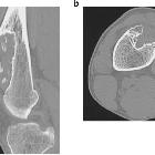
CT-Scan eines
periostalen Chondroms in distalen der Vorderfläche des Femurs. Beachte die Verkalkungen in der Matrix. Entnommen aus: Imura et al. 2014 A giant periosteal chondroma of the distal femur successfully reconstructed with synthetic bone grafts and a bioresorbable plate: a case report World Journal of Surgical Oncology. CC-BY 4.0 Licence

Lesions
involving the outer surface of the bone in children: a pictorial review. Parosteal osteosarcoma on the left distal posterior femur. A lateral radiograph of the left knee a shows a large exophytic mass with central dense ossifications adjacent to the femur (arrow). Sagittal T1 b and T2 c MR images demonstrate heterogeneous signal through this large soft-tissue tumor on both T1 and T2. A sagittal T1 post-contrast image d reveals heterogeneous contrast enhancement (arrow)

Lesions
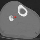
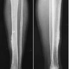
involving the outer surface of the bone in children: a pictorial review. Periosteal reaction post trauma. An antero-posterior radiograph of the leg bones a shows a healing fracture at the postero-medial aspect of the tibia. Adjacent cortical thickening and surrounding sclerosis are seen (arrow). Axial T2 b coronal T1 c and coronal T2 d images demonstrate a hypointense healing fracture line with the surrounding bone and soft-tissue edema

Lesions
involving the outer surface of the bone in children: a pictorial review. Ribbing disease. Lateral radiograph of right tibia a shows smooth thickening along the anterior cortex; this radiograph was obtained when the patient was 19 years of age. 7 years later, a follow-up lateral radiograph obtained as a young adult b shows further increased thickening along the anterior tibial cortex with narrowing of the medullary cavity. Axial c and sagittal d PD MR images of the tibia obtained at the same time as the second radiograph reveal smooth cortical thickening anteriorly. No unusual abnormal uptake of radiotracer was noted on the concomitantly performed Tc-99m MDP labeled bone scan e

Lesions
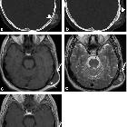
involving the outer surface of the bone in children: a pictorial review. Periosteal osteosarcoma in the right proximal femur. Axial a and coronal b T2 images reveal high signal soft-tissue mass involving predominantly the vastus medialis and intermedius with cortical disruption and intraosseous extension at the anteromedial aspect of the right femur (arrow)

Lesions
involving the outer surface of the bone in children: a pictorial review. Septic cortical osteitis. Antero-posterior radiograph of the left humerus a shows irregular inhomogenous lamellated periosteal reaction at the left lateral cortical surface of the diaphysis (thick arrow). There is an associated obliquely oriented fracture through the proximal humerus. Axial fat suppressed T2 b and coronal fat suppressed T2 c images demonstrate an intracortical hypointensity with surrounding edema through the medullary cavity and the adjacent soft tissues. An axial post-contrast injection image d shows peripheral contrast enhancement surrounding an intracortical hypointensity and enhancement through the medullary cavity and surrounding soft tissues (thin arrow)

Lesions
involving the outer surface of the bone in children: a pictorial review. Ganglion cyst at the lateral margin of the right knee in a 6-year-old boy. An AP radiograph of the right knee a reveals slight soft-tissue fullness along the lateral aspect of the knee (marked by star). This corresponded to the site of fluctuant mass noted on palpation. No associated adjacent bony defects/erosive changes are observed. Axial b and coronal c fat-suppressed T2-weighted MR images show a lobulated, deformable, cystic mass adjacent to and wrapping around the lateral distal femoral metaphysis (arrows). A sagittal proton density image d reveals high signal within the fluid (due to high mucopolysaccharide content); thin rim enhancement was noted with contrast (arrow) e

Lesions
involving the outer surface of the bone in children: a pictorial review. Trevor’s disease (dysplasia epiphysealis hemimelica) in 3-year-old boy. Antero-posterior radiograph of the left foot a shows asymmetric osteochondral overgrowth arising from the medial epiphysis of the tibia (arrow) and from multiple ossification centers within the tarsal bones of the foot. An axial STIR image b and axial and coronal T1 images c and d demonstrate cortico-medullary coalescence and cartilaginous signal (arrow)

Lesions
involving the outer surface of the bone in children: a pictorial review. A 5-year-old boy with bizarre parosteal osteochondromatous proliferation at the middle phalanx of the right index finger. Antero-posterior radiograph of the right index finger a demonstrates a well-defined mass with amorphous calcifications (arrow) adjacent to the otherwise intact cortical surface of the middle phalanx. No medullary continuity is demonstrated between the lesion and underlying bone. Axial T1 post-contrast image b shows irregular peripheral contrast enhancement of the lesion without surrounding soft-tissue invasion (arrow). The lesion is hypointense on coronal T1 c and mostly hyperintense with few hypointense foci on axial T2 d (black arrow)

Lesions
involving the outer surface of the bone in children: a pictorial review. An 11-year-old boy with juxta-cortical chondroma at the proximal right humerus. A radiograph of the left shoulder reveals mild scalloping at the medial cortex of the proximal humeral metaphysis (arrow). A coronal T2 STIR image b and a coronal T1 post-contrast image c show a soft-tissue lesion abutting the cortex with a heterogeneously high T2 signal intensity suggesting cartilage (thick arrow), and predominantly peripheral contrast enhancement (thin arrow)
Lesions
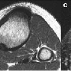
involving the outer surface of the bone in children: a pictorial review. Fibrous cortical defect. A 13-year-old boy with a fibrous cortical defect in the proximal right postero-medial tibia. An antero-posterior radiograph of right tibia a shows an intra-cortical lucency with a thin rim of sclerosis (arrow). Axial T1 b and T2 fat-suppressed c images show a hypointense intracortical lesion with no medullary involvement. A hyperintense rim is seen on the T2-weighted image (black arrow)

Lesions
involving the outer surface of the bone in children: a pictorial review. Benign solitary osteochondroma of the tibia in a 15-year-old boy. Lateral radiograph of the knee a shows a bony protrusion arising off the posterior aspect of the proximal tibial metaphysis; note the continuity with the medullary cavity of the underlying tibia (arrow). Sagittal b and axial c T2 MR images show a markedly hyperintense, thin cartilage cap surrounding this bony exostosis; an axial T1 post contrast MR image d reveals a thin, peripherally enhancing cartilage cap (arrow)

Periosteal
chondroma. Three-dimensional double-echo steady-state sequences: coronal
 Assoziationen und Differentialdiagnosen zu paraossale Tumoren:
bizarre
parosteale osteochondromatöse Proliferation


