perikardiale Ausläufer

Pericardial
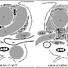
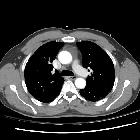
Recess: Computed Tomography Findings of Varying Disorders: Cross-section of the pericardial sinuses and recesses. The pericardial sinuses are composed of the TS and the OS. The TS is connected to the aortic recess (straight arrow), the right pulmonic recess (curved arrow), and the LPR (open arrow). The aortic recess is divided into the superior and the inferior aortic recesses. The SAR is the uppermost portion of the TS and extends upward along the right side of the ‘A’ usually to the level of the sternal angle. The SAR is subdivided into the anterior (single dot), the right lateral (two dots) and the posterior (three dots) sections. The LPR is located between the proximal portion of the right main ‘P’ and the LSPV, below the left main ‘P’. The OS lies behind the left atrium anterior to the ‘e’. The PPR of the OS is located posterior to the right ‘P’ and medial to the Br. The postcaval recess (*) behind the ‘S’ is a diverticulum of the pericardial cavity proper. The left pulmonic venous recess is located posterior to the LSPV. A = aorta, B = left main bronchus, Br = bronchus intermedius, e = esophagus, LPR = left pulmonic recess, LSPV = left superior pulmonary vein, OS = oblique sinus, P = pulmonary artery, PPR = posterior pericardial recess, RSPV = right superior pulmonary vein, S = superior vena cava, SAR = superior aortic recess, TS = transverse sinus

Pericardial
recesses • Pericardial recesses - Ganzer Fall bei Radiopaedia

Pericardial
recesses • Prominent pericardial recess on trauma CT - Ganzer Fall bei Radiopaedia

Pericardial
recesses • Superior periaortic pericardial recess - Ganzer Fall bei Radiopaedia
The pericardial recesses are small spaces in the pericardial cavity arising from the transverse pericardial sinus that are formed by the reflections of the pericardium. Pericardial fluid can pool in these recesses, mimicking mediastinal lymph nodes or pathology. There are several pericardial recesses that may be mistaken for dissection or lymphadenopathy:
- aortic recesses
- pulmonic recesses
- postcaval recess
- pulmonary venous recesses
Siehe auch:

 Assoziationen und Differentialdiagnosen zu perikardiale Ausläufer:


