Perikarderguss


Pericardial effusions occur when excess fluid collects in the pericardial space (a normal pericardial sac contains approximately 30-50 mL of fluid).
Epidemiology
There is no single demographic affected, as there are many underlying causes of pericardial effusion.
Clinical presentation
Clinical presentation of pericardial effusions does not relate so much to the size of the effusion but rather the speed at which the fluid has accumulated, as slow gradual accumulation allows the pericardium to stretch and accommodate much larger volumes of fluid .
Regardless of volume, symptoms relate to impaired cardiac function due to intrapericardial pressure approximating intracardiac pressure leading to an impaired filling of low-pressure chambers, particularly the right atrium.
Dyspnea and reduced exercise tolerance will be early signs, progressing to severe impaired cardiac output and death in severe cases (e.g. cardiac tamponade).
Pathology
Etiology
- idiopathic (presumed viral, post-viral or immune-related)
- inflammatory
- systemic inflammatory conditions or connective tissue diseases
- post-myocardial infarction: Dressler syndrome
- infectious
- viral
- bacterial
- tuberculosis
- post-surgical or traumatic
- pulmonary arterial hypertension
- radiotherapy
- malignancy
- endocrine
Radiographic features
Plain radiograph
Small pericardial effusions are often occult on plain film. Greater than 200 mL of pericardial fluid is usually required to become radiographically visible. Radiographic signs include:
- there can be globular enlargement of the cardiac shadow giving a water bottle configuration
- lateral CXR may show a vertical opaque line (pericardial fluid) separating a vertical lucent line directly behind the sternum (pericardial fat) anteriorly from a similar lucent vertical lucent line (epicardial fat) posteriorly; this is known as the Oreo cookie sign
- widening of the subcarinal angle without other evidence of left atrial enlargement may be an indirect clue
- a differential density sign at cardiac borders has been suggested , but its specificity is limited
- serially enlarging cardiothoracic ratio
- hemodynamic compromise may manifest with signs of cardiogenic pulmonary edema
Echocardiography
Echocardiography is the method of choice to confirm the diagnosis, estimate the volume of fluid and most importantly assess the hemodynamic impact of the effusion.
An anechoic separation of pericardial layers can be identified on transthoracic echocardiography when pericardial fluid exceeds 50 mL. In a supine patient, the effusion will first appear posteriorly, tracking anterior to the descending aorta and left atrium. The parasternal long-axis and subcostal four-chamber views are typically favored for inspection of the pericardial space.
Differentiation from an epicardial fat pad and a left pleural effusion, which have similar sonographic appearances, relies on the recognition of the anatomical boundaries of the fluid collection in question; pleural effusions are bounded anteriorly by the descending aorta, and a fat pad will be seen most prominently in the atrioventricular groove. Additionally, epicardial fat pads have a different echotexture, with low-level echoes scattered throughout .
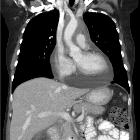
CT
The accepted thickness of a normal pericardium, measured on CT scans and on MR images, is often taken at 2 mm and abnormal is considered >3-4 mm .
CT makes the diagnosis extremely easy but is usually obtained to try and clarify the cause of an effusion rather than to confirm the diagnosis. Pericardial effusions are a frequent incidental finding in unwell hospitalized patients.
Fluid density material is seen surrounding the heart. Careful inspection of the region is necessary to ensure that no invasive mass can be identified.
Estimating volume
The depth of the effusion can be used to estimate the likely volume of fluid, provided the fluid is relatively evenly spread throughout the pericardium (i.e. global effusion) . Clearly, this does not apply to loculated effusions.
- <5 mm: 50-100 mL
- 5-10 mm: 100-250 mL
- 10-20 mm: 250-500 mL
- >20 mm: >500 mL
However, due to complex pericardial anatomy and fluid being able to pool in the pericardial recesses this relationship is not exact, and it may be better to report volume in more general terms :
- <10 mm: small
- 10-20 mm: moderate
- >20 mm: large
Treatment and prognosis
If small, asymptomatic and clinically not-suspect then conservative management is usually favored.
If large, symptomatic or there is a clinical concern of the underlying cause (e.g. infection, malignancy) then pericardiocentesis can be performed to drain the fluid. A Seldinger technique is employed, usually under ultrasound/echocardiographic guidance, to insert a drain into the pericardial space .
In cases where effusions are recurrent and symptomatic (e.g. malignancy) then pericardial fenestration can be performed.
Differential diagnosis
- hemopericardium: has higher attenuation on CT and often a different clinical context
- cardiomegaly: can sometimes mimic an effusion
See also
Siehe auch:
- Kardiomegalie
- hämorrhagischer Perikarderguss
- fetal pericardial effusion
- eitrige Perikarditis
- Perikarddrainage
- Herzbeuteltamponade
- pericardiotomy
und weiter:
- Herzkonfiguration
- cardiothoracic ratio
- obstetric curriculum
- Hydrops fetalis
- Perikardempyem
- Ebstein anomaly
- mediastinal lymphoma
- enlargement of the cardiac silhouette
- rheumatic heart disease
- Perikarditis
- Dilatative Kardiomyopathie
- water bottle sign
- cardiac curriculum
- large-cell lymphoma of the mediastium
- kardiale Metastasen
- cardiovascular manifestations of AIDS
- Migrating pericardial cyst
- Myokardruptur
- exsudative Perikarditis
- response evaluation criteria in solid tumours
- oreo cookie sign
- Chyloperikard
- perikardiale Ausläufer
- PE - als Abkürzung


 Assoziationen und Differentialdiagnosen zu Perikarderguss:


