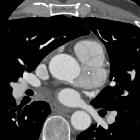
Primary cardiac tumors

Großes
Vorhofmyxom mit Ausdehnung vor allem in den linken Vorhof und hämodynamischer Wirkung, so dass es zu einer beginnenden Dekompensation kam. Siehe auch Pleuraergüsse rechts mehr als links. Die Dichtewerte in der Computertomographie schwankten um ca. 25 bis 30 HE.

Significant
incidental cardiac disease on thoracic CT: what the general radiologist needs to know. Axial contrast-enhanced CT demonstrates a mass (black arrows) involving the right atrium and ventricle, (later confirmed to be angiosarcoma by histology) on axial contrast enhanced CT and reformatted images in a 27-year-old female being investigated for possible pulmonary embolism (shortness of breath and dizziness)

Primary
cardiac sarcoma: reports of two cases and a review of current literature. CT scan shows a large filling defect (black arrow) in the left atrium.

Complete
resection of undifferentiated cardiac sarcoma and reconstruction of the atria and the superior vena cava: case report. Cardiac magnetic resonance imaging demonstrating a mass in the right atrium involving the left atrial dome and atrial septum.

Left-sided
primary cardiac lymphoma: a case report. Transthoracic echocardiography showing the atrial mass, long axis

Left-sided
primary cardiac lymphoma: a case report. CT image of the atrial mass and the mediastinal bulky

Left-sided
primary cardiac lymphoma: a case report. MRI image of the atrial mass and the mediastinal bulky

Primary
pericardial mesothelioma. Coronal reformatted CT image demonstrating multiple enlarged lymph nodes. Liver cyst.

Primary
cardiac tumors • Cardiac hemangioma - Ganzer Fall bei Radiopaedia

Primary
cardiac tumors • Cardiac tumor - undifferentiated pleomorphic sarcoma - Ganzer Fall bei Radiopaedia

Primary
cardiac tumors • Cardiac angiosarcoma - Ganzer Fall bei Radiopaedia

Primary
cardiac tumors • Cardiac paraganglioma - Ganzer Fall bei Radiopaedia
Primary cardiac tumors are uncommon and comprise only a small minority of all tumors that involve the heart: most are mediastinal or lung tumors that extend through the pericardium and into the heart, or metastases .
Epidemiology
Primary cardiac tumors have an estimated autopsy prevalence of 0.001-0.03% .
Pathology
Primary cardiac tumors can then be divided into:
- benign cardiac tumors: 60-75%
- cardiac myxoma: most common in adults
- cardiac lipoma (≈10% , second most common in adults )
- cardiac rhabdomyoma: most common in children
- papillary fibroelastoma
- cardiac fibroma
- cardiac hemangioma
- cardiac paraganglioma
- pericardial teratoma (can rapidly grow despite being benign)
- malignant cardiac tumors
- sarcomas account for 25% of all cardiac tumors
- cardiac angiosarcoma: most common malignant primary cardiac tumor
- undifferentiated sarcoma of the heart
- cardiac leiomyosarcoma
- cardiac spindle cell sarcoma
- cardiac fibrosarcoma
- cardiac liposarcoma
- primary cardiac osteosarcoma: 3-9% of primary cardiac malignant tumors
- malignant fibrous histiocytoma of heart
- cardiac hemangiopericytoma
- primary cardiac lymphoma
- pericardial mesothelioma
- sarcomas account for 25% of all cardiac tumors
Siehe auch:
- Lipomatöse Hypertrophie des interatrialen Septums
- Vorhofmyxom
- intrakardiale Thromben
- papilläres Fibroelastom des Herzens
- Rhabdomyom des Herzens
- Angiosarkom des Herzens
- kardiale Metastasen
- maligne Tumoren des Herzen
- kardiales Hämangiom
- Myxom des linken Vorhofs
- kardiales Fibrom
- kardiales Lipom
- benigne Tumoren des Herzens
- primäres Mesotheliom des Perikards
- fetales perikardiales Teratom
- primäres kardiales Lymphom
- Crista terminalis atrii dextri
und weiter:

 Assoziationen und Differentialdiagnosen zu primäre kardiale Tumoren:


