Angiosarkom des Herzens

Primary
cardiac metastatic angiosarcoma. Images demonstrate the heterogeneous signal intensity intracardiac mass connected to the posterolateral wall of the right atrium, which appears to be infiltrated (arrows), with signs of extra-auricular extension. There is also pleural effusion (asterisks).

Significant
incidental cardiac disease on thoracic CT: what the general radiologist needs to know. Axial contrast-enhanced CT demonstrates a mass (black arrows) involving the right atrium and ventricle, (later confirmed to be angiosarcoma by histology) on axial contrast enhanced CT and reformatted images in a 27-year-old female being investigated for possible pulmonary embolism (shortness of breath and dizziness)

Unusual CT
features of primary cardiac metastatic angiosarcoma. Anterior mediastinal tumour (asterisk), with irregular borders (arrows).

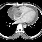
Unusual CT
features of primary cardiac metastatic angiosarcoma. The tumour (asterisk) is in close proximity with the right atrium, as its free border cannot be depicted.

Primary
cardiac tumors • Cardiac angiosarcoma - Ganzer Fall bei Radiopaedia
Angiosarcoma
• Right atrial angiosarcoma - Ganzer Fall bei Radiopaedia
Cardiac angiosarcomas are the most common sarcoma involving the heart (see cardiac tumors).
Please refer to the article on angiosarcomas for a general discussion about this entity.
Epidemiology
They occur slightly more frequently in males.
Clinical presentation
Patients usually present with right-sided heart failure or cardiac tamponade.
Pathology
These tumors tend to occur in the right atrium and involve the pericardium.
Radiographic features
Two main morphologic types have been described in angiosarcoma:
- CT shows a low-attenuation right atrial mass, which may be irregular or nodular (usually arises from the right atrial free wall)
- contrast material enhancement is heterogeneous
- pericardial space may be obliterated with hemorrhagic, necrotic tumor debris
Treatment and prognosis
Prognosis is poor, which may be due in part to the delay in diagnosis (patients usually have metastatic involvement at presentation).
Siehe auch:
- Angiosarkom
- Tumoren des Herzens
- Kaposisarkom
- primäre maligne Neoplasien des Herzens
- HIV associated neoplasms
- Tumoren des rechten Vorhofs
und weiter:


 Assoziationen und Differentialdiagnosen zu Angiosarkom des Herzens:
