Perikardzyste


Pericardial cysts are uncommon benign congenital anomalies of the anterior and middle mediastinum.
Clinical presentation
Usually asymptomatic and discovered incidentally although occasionally may present with chest pain and dyspnea.
Pathology
They are thought to often result from aberrations in the formation of celomic cavities. They can occur as sequelae of previous pericarditis. The cyst wall is composed of connective tissue and a single layer of mesothelial cells, and usually contains clear fluid.
Location
They are most commonly found on the right side, in particular the right anterior cardiophrenic angle, but can be found almost anywhere adjacent to the heart.
Variants
Radiographic features
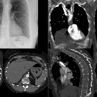
Plain radiograph
Typically seen as a mass-like density at the cardiophrenic sulcus. They can be of different shapes and are not always round. May change in shape and size with inspiration and position.
CT
Usually appears as a well-defined, non-enhancing, fluid-attenuation, rounded mass next to the pericardium.
MRI
Morphology again can be variable. Internal septations may be present. Signal characteristics are those of fluid and include :
- T1: typically low signal (occasionally can be high signal if contains proteinaceous material)
- T2: high signal
- T1 C+ (Gd): no enhancement
Treatment and prognosis
They are benign lesions. Surgical resection or aspiration may be performed for symptomatic selected cases.
Differential diagnosis
As general differential on cross-sectional imaging
On a chest radiograph also consider:
Siehe auch:
- Bronchogene Zyste
- cystic mediastinal masses
- mediastinales Teratom
- Morgagni-Hernie
- Perikarditis
- Tumoren des Thymus
- Tumoren des Perikards
- perikardiale Ausläufer
und weiter:


 Assoziationen und Differentialdiagnosen zu Perikardzyste: