Duplikationszyste des Vorderdarms

Peritoneal
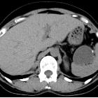
metastatic adenocarcinoma possibly due to a gastric duplication cyst: a case report and literature review. Computed tomography of the abdomen (with contrast agent). A well-defined oval lesion is seen immediately adjacent to the gastric corpus and left adrenal gland area. A linear septum can also be seen.

Successful
laparoscopic resection for gastric duplication cyst: a case report. Abdominal ultrasound revealing a cystic lesion with a clearly defined boundary of approximately 40 mm in the pancreatic tail
Successful
laparoscopic resection for gastric duplication cyst: a case report. A thick cystic lesion of the septum is visible in the pancreatic tail, but computed tomographic scan shows no invasion into the stomach wall

Successful
laparoscopic resection for gastric duplication cyst: a case report. Endoscopic ultrasound showing the tumor, which appears smooth with a marginal edge, characterized by echo with high homogeneity, and the presence of viscous mucus was suspected

Cystic liver
lesions: a pictorial review. Ciliated hepatic foregut duplication cyst in a 15-year-old male. a Axial non-enhanced computed tomography shows a subcapsular nodule of the fourth segment with spontaneous attenuation value of around 50 Hounsfield Units. b On axial T1-weighted magnetic resonance imaging, it is spontaneously hyperintense. c It displays a high level of hyperintensity on axial T2-weighted magnetic resonance imaging. d Axial T1-weighted magnetic resonance imaging on portal venous phase shows neither contrast enhancement nor wall thickening

Foregut
duplication cyst • Foregut duplication cyst - Ganzer Fall bei Radiopaedia

Foregut
duplication cyst • Foregut duplication cyst - Ganzer Fall bei Radiopaedia

Foregut
duplication cyst • Foregut duplication cyst - Ganzer Fall bei Radiopaedia

Foregut
duplication cyst • Superior mediastinal mass due to esophageal duplication cyst - Ganzer Fall bei Radiopaedia

Foregut
duplication cyst • Appendicitis and incidental foregut duplication cyst - Ganzer Fall bei Radiopaedia

Foregut
duplication cyst • Foregut duplication cyst with vertebral anomalies - Ganzer Fall bei Radiopaedia
Foregut duplication cysts are a type of congenital duplication cyst. They are sometimes classified under bronchopulmonary foregut malformations.
Entities classified as foregut duplication cysts include:
- bronchogenic cysts
- neurenteric cysts
- other enteric cysts
- esophageal duplication cysts
- lingual duplication cysts - rare
- gastric duplication cysts - rare
See also
Siehe auch:
- Lungensequester
- Bronchogene Zyste
- Hämangiom
- Perikardzyste
- Duplikationszyste des Magens
- Duplikationszysten gastrointestinal
- Duplikationszyste des Ösophagus
- neuroenterische Zyste
- benignes Ganglioneurom
und weiter:


 Assoziationen und Differentialdiagnosen zu Duplikationszyste des Vorderdarms:


