Phlebolithen in skrotaler Varicocele

Phlebolithen in Varikozele
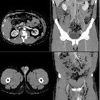
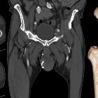
Phlebolithen in skrotaler Varicocele
Phlebolithen Radiopaedia • CC-by-nc-sa 3.0 • de
Phleboliths are literally "vein stones", and represent calcification within venous structures. They are particularly common in the pelvis where they may mimic ureteric calculi, and are also encountered frequently in venous malformations. There is an association with Maffucci syndrome.
Radiographic features
Phleboliths appear as focal calcifications, often with radiolucent centers (if present, a helpful sign to distinguish them from urolithiasis). This appearance is attributed to calcification peripherally within the vessel and is frequently seen on abdominal radiographs (66% of phleboliths ). It can also be seen on CT provided thin sections are obtained (at 5 mm thick slices radiolucent centers will be inapparent in 99% of phleboliths ).
Differential diagnosis
Two signs are helpful in distinguishing a ureteric calculus from a phlebolith:
Siehe auch:


 Assoziationen und Differentialdiagnosen zu Phlebolithen in Varikozele: