soft tissue venous malformations


Soft tissue venous malformations, commonly known as soft tissue hemangiomas, are a location-dependent benign vascular soft tissue tumor.
They are the most common angiomatous lesions and represent up to 7% of all benign soft-tissue tumors .
Terminology
It is important to note that according to newer nomenclature (ISSVA classification of vascular anomalies), these lesions are merely known as slow flow venous malformations. Having said that, it is probably helpful in reports to include the word 'hemangioma' as this term is ubiquitous in the literature and most familiar to many clinicians. The remainder of this article uses the terms 'soft tissue hemangioma' and 'soft tissue venous malformation' interchangeably.
Epidemiology
There may be a greater female predilection. In the pediatric population, hemangiomas tend to be the most frequently diagnosed soft-tissue neoplasm.
Pathology
Subtypes
Soft tissue hemangiomas may be classified into five histological subtypes.
This classification is dependent on the predominant type of vascular channel identified within them:
- capillary: commonest type; tend to predominate in the pediatric population.
- cavernous
- arteriovenous
- venous
- mixed
Location
They can arise in various anatomic locations, including striated muscle, skin, subcutaneous tissue, and synovial tissue (synovial hemangioma).
Radiographic features
Plain radiograph
Small lesions may be occult on plain film, while large lesions may show evidence of a focal soft tissue swelling +/- associated phleboliths.
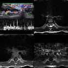
Ultrasound
Can have a variable appearance. Typically seen as an ill-defined or well-defined hypoechoic mass of heterogeneous echotexture with multiple cystic spaces within. On color Doppler, there may be no detectable signal or only weak signal .
CT
On unenhanced CT, it may appear as an ill-defined mass of similar attenuation to muscle. CT may also show the presence of associated phleboliths.
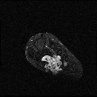
MRI
Hemangiomas are typically well-defined, lobulated and heterogeneous with no features of local invasion.
While many sequences show a rather heterogeneous signal mass certain signal characteristics tend to dominate.
- T1
- overall signal is often intermediate to slightly high (relative to skeletal muscle)
- some focal high signal areas may be present in a large proportion of lesions (up to 70% )
- T2: high signal intensity tends to dominate on T2-weighted images
- gradient echo: the presence of phleboliths may show blooming artifact
- T1 C+ (Gd): lesions show marked signal enhancement in parts of the areas, which were both of high and low T2
Some intramuscular hemangiomas may also be associated with atrophic changes in muscles.
Siehe auch:
- Phlebolithen
- Hämangiom
- WHO Klassifikation der Weichteiltumoren
- venöse Malformationen
- intramuskuläre Verkalkungen
- low flow vascular malformation
- multiple haemangiomas of the head and neck
- synoviales Hämangiohamartom
- venöse Malformationen des Gesichts
und weiter:


 Assoziationen und Differentialdiagnosen zu Weichteilhämangiom:


