Pseudocyst of the humerus


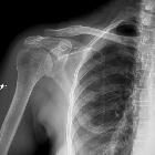

Pseudocyst of the humerus, also referred as a humeral head pseudolesion, is a normal anatomical variant due to increased cancellous bone in the region of the greater tuberosity of the humerus which is seen as a lucent lesion on radiography.
Hyperemia and disuse caused by shoulder problems (such as rotator cuff disorders) cause this area of lucency to appear more lucent and mimic a lytic lesion (e.g. chondroblastoma, infection, or even a metastatic focus) and mistakenly undergo biopsy.
Therefore, pseudocyst of the humerus is one of the skeletal "do not touch" lesions.
Radiographic features
Plain radiograph
May show a lucent lesion in the region of the humeral greater tuberosity, often with a somewhat well defined inferior curvilinear margin whilst the upper margin is generally ill-defined . Pseudocysts are thought to be so radiographically characteristic that they should not be biopsied.
CT
Similar appearances to those on plain radiograph.
Nuclear medicine - bone scan
Bone scan may show increased radionuclide uptake due to hyperemia (and thus convince the surgeon to perform a biopsy).
Siehe auch:
und weiter:


 Assoziationen und Differentialdiagnosen zu Pseudozyste des Humeruskopfes: