Quadriceps injuries


Quadriceps injuries are injuries affecting the quadriceps muscle or quadriceps tendon and comprise a spectrum of strains, tears, avulsion and contusions up to the quadriceps tendon rupture.
Epidemiology
Quadriceps injuries are common injuries in athletes and the quadriceps muscle is often affected by muscle strains in situations requiring explosive movements as seen in certain sports .
The rectus femoris muscle is the most commonly involved in a quadriceps injury .
Vastus muscle injury seems to be less common, the vastus intermedius muscle seems to be most commonly affected by contusions .
Quadriceps tendinosis is a typical overuse injury seen in athletes with an estimated prevalence of 0.2-2% in that population .
Quadriceps tendon rupture is a rather uncommon but important and debilitating diagnosis, occurring more often in older men .
Clinical presentation
Most muscle injuries can be elucidated by a thorough history and physical examination. Symptoms will vary with the type and location of the injury.
Quadriceps muscle contusions can be easily elucidated by a history of blunt trauma and clinical examination will usually reveal skin discolouration, tenderness, swelling and varying degrees of pain and tenderness as well as limited range of motion and difficulties to weight bear.
Quadriceps muscle strains might reveal a more variable clinical history ranging from a sharp thigh pain and/or hip pain associated with movements to vague discomfort or thigh enlargement and variably associated strength deficit . Tenderness upon direct palpation of the muscle strain is a typical finding and pain can usually be triggered by stretching and resisted muscle activity. Palpable defects are another clinical finding, which can be used for clinical grading together with the amount of pain and strength deficit associated with the injury .
Quadriceps tendinosis presents as pain at the superior patellar pole aggravated by knee flexion.
In the case of quadriceps tendon rupture, patients will present with pain and difficulties to weight bear. The physical examination might reveal a palpable defect above the superior patella pole and patients are not able to extend the knee against resistance or to perform a straight leg raise.
Complications
Complications will vary with the type of injury and include the following :
- progression to complete tear or tendon rupture
- re-injury
- muscle atrophy and fatty replacement
- compartment syndrome (in case of crush injury)
- myositis ossificans
Pathology
Mechanism
The injury mechanism varies with the type of injury. Muscle contusions and crush injuries are caused by direct trauma . Proximal tendon tears or tendon avulsion injuries, myotendinous and myofascial strain injuries are usually caused by eccentric loading mechanisms. Quadriceps tendinosis is a typical overuse injury and quadriceps tendon rupture can be caused by both eccentric loading or direct impact .
Location
Most quadriceps tendon strain injuries concern the rectus femoris muscle. It crosses two joints, features fast-twitching (type II) fibers and complex musculotendinous anatomy and undergoes forceful eccentric contraction during sprinting, jumping and kicking . Direct trauma can affect any part of the quadriceps femoris, the vastus intermedius muscle seems the most commonly affected muscle . Quadriceps tendinosis and tendon rupture obviously affect the distal quadriceps tendon .
Subtypes
Quadriceps injuries can be classified based on type and location into the following:
- rectus femoris injury
- anterior inferior iliac spine avulsion
- proximal rectus femoris tendon injury
- myotendinous injury (common)
- myofascial injury (uncommon)
- vastus muscle injury
- quadriceps tendon injury
- muscle contusion
- muscle laceration
Radiographic features
The diagnosis of a quadriceps injury can be usually made clinically with a thorough history and physical examination . Imaging and in particular ultrasound and MRI can provide information regarding the extent, type and prognosis of the muscle injury .
Plain radiograph
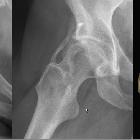
Plain radiographs pelvis can be used as an initial examination to visualize avulsions to rule out fractures and other pathology.
Ultrasound
The quadriceps muscle can be easily assessed by ultrasound, which can nicely depict the proximal rectus femoris tendons, the rectus femoris muscle and the vastus muscles. The quadriceps tendon can be best visualized when the knee is flexed .
CT
CT can detect and characterize avulsion injuries. Additionally, it can be of used to depict large intramuscular hemorrhage. Due to radiation exposure and alternatives with better soft tissue discrimination properties as ultrasound and MRI, its value in the workup of muscle injuries is limited and might be used only in case of crush injuries or polytraumatized patients for the workup of associated injuries.
MRI
Typical features of muscle injuries on MRI include fluid signal intensity tracking along and surrounding the muscle fibers, myofascial, myotendinous or tendinous units and/or discontinuities of the injured muscle. Higher grade injuries might include tendon and muscle retraction .
Depending on the type and location of injury appearances will vary. Direct impact injuries are more often associated with hematomas .
An MR based classification is the British Athletics muscle injury classification, which also contains a subclassification according to the site.
Radiology report
The radiological report should include a description of the following:
- location type and extent of the lesion
- the extent of tendon retraction
- associated injuries
Treatment and prognosis
Management of quadriceps injuries will depend on the type and location of the injury. A complete quadriceps tendon tear with loss of the extensor mechanism requires surgery and reattachment of the quadriceps tendon.
Most other quadriceps injuries are managed conservatively and a currently accepted method to treat muscle strain injuries in the initial period follows the RICE (rest, ice, compression and limb elevation) principle. An initial resting period serves as a measure to prevent further progression of the injury and a more severe strain injury might require an initial use of crutches. Limb elevation and intermittent application of ice and compression are aiming to decrease blood flow and increased amounts of interstitial fluid accumulation. Ice application also serves pain control and can be supplemented by non-steroidal anti-inflammatory drugs (NSAIDs) in the initial period .
After the initial phase (usually 3-5 days), treatment should be followed up by a rehabilitation protocol comprising movement, walking, running exercises as well as stretching, strengthening, range of motion, endurance and agility training. Exercises should be initiated gradually and advanced continuously and should be conducted without increasing pain in the quadriceps .
Muscle contusions of the quadriceps are managed similar to muscle strain injuries except that the injured leg should be placed in a flexed position for the first 24 hours and compression should be kept to prevent hematoma formation (e.g. hinged knee brace or compression wrap). Similar to muscle strains cryotherapy should be administered .
Recovery will depend on the type and extent of the injury and might take from 2-3 weeks in case of a muscle contusion or myofascial injury and up to 4 months or longer in a tendon avulsion or tendon rupture .
Differential diagnosis
Important differential diagnosis of quadriceps injuries are the following :
See also

 Assoziationen und Differentialdiagnosen zu Ruptur Musculus quadriceps femoris:
