Rosette-forming glioneuronal tumors
 nicht verwechseln mit: Embryonaler Tumor mit mehrreihigen Rosetten
Rosette-forming glioneuronal tumors (RGNTs) are rare, usually midline, tumors that involve the fourth ventricle and/or aqueduct of Sylvius.
Although relatively well circumscribed on MRI and clinically indolent, they often invade surrounding tissues, involving the cerebellum, pons and even the pineal region. There are often cystic components and they tend to have heterogeneous contrast enhancement. They are considered WHO grade I lesions in the current (2016) WHO classification of CNS tumors.
Epidemiology
Rosette-forming glioneuronal tumors are typically diagnosed in younger adult patients with a mean age of 30 years .
Clinical presentation
Symptoms are non-specific and mainly depend on the location of the tumor. Commonly they present with ataxia and headache.
Pathology
Location
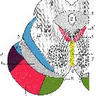
Rosette-forming glioneuronal tumors are originally described in the region of the 4th ventricle (~60%) often with variable local parenchymal extension .A recent literature review has demonstrated these tumors outside the characteristic location including the cerebellum hemispheres, cerebellopontine angle, pineal gland, midbrain tectum, thalamus, third ventricle, optic chiasm and spinal cord.
Microscopic features
Rosette-forming glioneuronal tumors contain both neurocytic and astrocytic components. The neurocytic component, which is a minor component but allows for diagnosis of this tumor, forms perivascular pseudorosettes or neurocytic rosettes surrounding eosinophilic neuropil cores . The larger glial component resembles that of pilocytic astrocytomas, including the presence of Rosenthal fibers .
Immunophenotype
Immunohistochemistry is consistent with the biphasic nature of this tumor with both glial and neurocytic patterns identified.
The glial component demonstrates:
The neurocytic component demonstrates:
- MAP2: positive
- neuron-specific enolase: positive
- synaptophysin: positive in the center of the neurocytic rosettes
Proliferation index (Ki-67) is usually low, <3% .
Radiographic features
Rosette-forming glioneuronal tumors have variable solid-cystic components :
- entirely solid: 40%
- mixed solid and cystic: 35%
- entirely cystic: 25%
CT
On CT these tumors are of variable appearance reflecting the solid and cystic components. Calcification is seen in approximately a quarter of cases .
MRI
As expected, the cystic components demonstrate high T2 signal, whereas the solid components demonstrate variable gadolinium enhancement .
Treatment and prognosis
If complete resection is achievable, which depends mostly on the location of the tumor, then a cure is possible to surgery alone.
Differential diagnosis
The main differential diagnosis is pilocytic astrocytoma. The other differential diagnoses will, to a degree, depend on the location and include:
- pilocytic astrocytoma
- gangliogliomas and gangliocytomas
- pleomorphic xanthoastrocytoma (almost always hemispheric)
- papillary glioneuronal tumor (usually hemispheric)
Siehe auch:
- Pons
- Cerebellum
- Grundlagen der Magnetresonanz-Tomographie
- vierter Ventrikel
- Embryonaler Tumor mit mehrreihigen Rosetten
- DNETs
- glioneuronal tumours
- pineal region
und weiter:


 Assoziationen und Differentialdiagnosen zu rosette-forming glioneuronal tumour of the fourth ventricle: