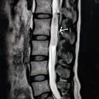
Schwannom der Cauda equina

Coexistence
of intervertebral disc herniation with intradural schwannoma in a lumbar segment: a case report. T2-weighted sagittal image (a) revealed L3/4 disc herniation which compressed the dural sac from the right side. Two slices away, there was a hyperintense intradural mass at the same level (b). T1-weighted sagittal image (c) demonstrated a hypointense mass behind the L3/4 intervertebral disc

Coexistence
of intervertebral disc herniation with intradural schwannoma in a lumbar segment: a case report. Gadolinium contrast MR images (a coronal image, b sagittal image) demonstrated a partially enhanced intradural mass at the left side of the L3/4 spinal canal. Axial image (c) showed an intradural mass of heterogeneous signal at the left and a herniated disc at the right extradural space
 Assoziationen und Differentialdiagnosen zu Schwannom der Cauda equina:


